. 2008 Nov;190(22):7523-31.
doi: 10.1128/JB.00945-08. Epub 2008 Sep 19.

SciN is an outer membrane lipoprotein required for type VI secretion in enteroaggregative Escherichia coli

Affiliations

SciN is an outer membrane lipoprotein required for type VI secretion in enteroaggregative Escherichia coli

Marie-Stéphanie Aschtgen et al. J Bacteriol. 2008 Nov.

Abstract

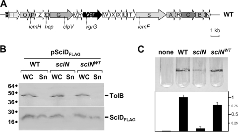
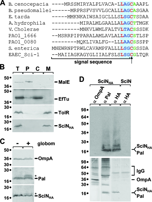
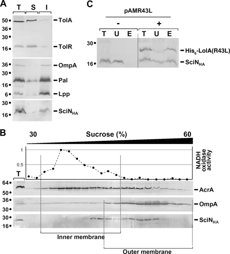
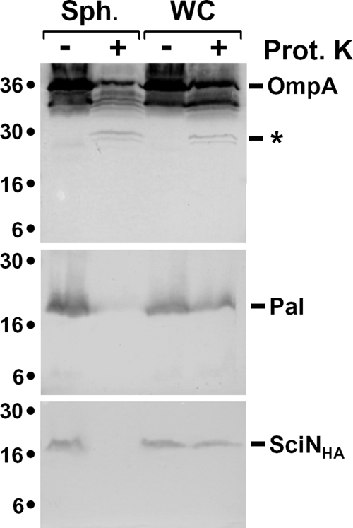
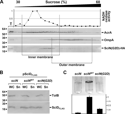
Enteroaggregative Escherichia coli (EAEC) is a pathogen implicated in several infant diarrhea or diarrheal outbreaks in areas of endemicity. Although multiple genes involved in EAEC pathogenesis have been identified, the overall mechanism of virulence is not well understood. Recently, a novel secretion system, called type VI secretion (T6S) system (T6SS), has been identified in EAEC and most animal or plant gram-negative pathogens. T6SSs are multicomponent cell envelope machines responsible for the secretion of at least two putative substrates, Hcp and VgrG. In EAEC, two copies of T6S gene clusters, called sci-1 and sci-2, are present on the pheU pathogenicity island. In this study, we focused our work on the sci-1 gene cluster. The Sci-1 apparatus is probably composed of all, or a subset of, the 21 gene products encoded on the cluster. Among these subunits, some are shared by all T6SSs identified to date, including a ClpV-type AAA(+) ATPase (SciG) and an IcmF (SciS) and an IcmH (SciP) homologue, as well as a putative lipoprotein (SciN). In this study, we demonstrate that sciN is a critical gene necessary for T6S-dependent secretion of the Hcp-like SciD protein and for biofilm formation. We further show that SciN is a lipoprotein, as shown by the inhibition of its processing by globomycin and in vivo labeling with [(3)H]palmitic acid. SciN is tethered to the outer membrane and exposed in the periplasm. Sequestration of SciN at the inner membrane by targeting the +2 residue responsible for lipoprotein localization (Gly2Asp) fails to complement an sciN mutant for SciD secretion and biofilm formation. Together, these results support a model in which SciN is an outer membrane lipoprotein exposed in the periplasm and essential for the Sci-1 apparatus function.


Figures

FIG. 1.
FIG. 1.
SciN is required for T6S. (A) Schematic organization of the EAEC sci-1 gene cluster. Conserved genes are indicated by gradations of gray to black. White genes are not conserved among T6S gene clusters. The generic names of conserved genes characterized are indicated below. (B) Effect of the sciN mutation on Hcp-like SciD protein secretion. SciD-FLAG secretion was assessed by separating whole cells (WC) and supernatant (Sn) fractions from WT, sciN, and complemented sciN (sciNWT) strain cultures. A total of 2 × 10⁸ cells and the TCA-precipitated material of the supernatant from 5 × 10⁸ cells were loaded on a 15% acrylamide SDS-PAGE gel and subjected to immunodetection using anti-FLAG monoclonal antibody (lower panel) and the anti-TolB polyclonal antibodies (upper panel). (C) Effect of the sciN mutation on biofilm formation. Biofilms formed in static cultures of WT, sciN, and complemented sciN cells (none, LB medium) were visualized in glass tubes by crystal violet staining (upper panel) and quantified using the ethanol-solubilization procedure, relative to the WT EAEC strain (lower graph).
FIG. 2.
FIG. 2.
SciN is a lipoprotein. (A) Sequence alignment using Clustal W of the N terminus of the SciN protein from the EAEC Sci-1 T6SS and homologous genes (COG3521 family) from T6SS of Burkholderia pseudomallei strain K96248 (YP_112106), E. tarda (ABW69084), Aeromonas hydrophila strain ATCC7966 (YP_856372), V. cholerae strain V52 (ZP_01680158), P. aeruginosa strain PAO1 HSI-1 (PA0080; NP_248770) and HSI-2 (PA1666; NP_250357), S. enterica serovar Typhi strain CT18 (NP_454883), and B. cenocepacia strain HI2424 (YP_834112). The putative lipobox motif, L-G/A/SG/A/S-C, is indicated by the blue box with the invariable residues in red. The positions of the signal sequence and the acylated N-terminal cysteine residue (arrow) are indicated. The residue in green corresponds to the +2 residue, which is responsible for the final localization of the lipoprotein in the cell envelope. (B) SciN cofractionates with membranes. A fractionation procedure was applied to EAEC cells producing SciN-HA (lane T), allowing separation between the periplasmic (lane P), cytoplasmic (lane C), and membrane fractions (lane M). Samples were loaded on a 15% acrylamide SDS-PAGE gel and subjected to immunodetection with antibodies directed against the periplasmic MalE, the cytoplasmic EfTu, the membrane TolR proteins, and the HA epitope (from top to bottom panel). Molecular weight markers are indicated on the left. (C) SciN is not processed in the presence of globomycin. A total of 2 × 10⁸ EAEC cells producing the HA-tagged SciN protein treated (+) or not (−) by the SPII inhibitor antibiotic globomycin (globom) were loaded on a 15% acrylamide SDS-PAGE gel and subjected to immunodetection by anti-OmpA, anti-Pal, or anti-HA antibody (from top to bottom panel). The unprocessed species of the Pal lipoprotein and SciN-HA are indicated by asterisks. Molecular weight markers are shown on the left. (D) SciN is labeled by [3H]palmitate. A total of 3 × 10⁹ cells of EAEC producing SciN or SciN-HA were labeled in vivo by [3H]palmitic acid, SDS-Triton X-100 solubilized, and immunoprecipitated using anti-OmpA, anti-Pal, and anti-HA antibodies, as indicated. Samples were loaded on 15% acrylamide SDS-PAGE gels and subjected to autoradiography (upper panel) or immunodetection by corresponding antibodies (lower panels). Molecular weight markers are indicated on the left. α, anti; IgG, immunoglobulin G.
FIG. 3.
FIG. 3.
SciN is an OM protein. (A) SciN is not solubilized by SLS. Total (T) membranes from EAEC cells producing SciN-HA were treated with SLS, and solubilized (S) and insolubilized (I) membranes were separated. Samples from 5 × 10⁸ cells were loaded on 15% acrylamide SDS-PAGE gels and subjected to immunodetection with antibodies directed against TolA, TolR (upper panel), and Pal (which recognizes OmpA and Lpp; middle panel) (11) and against the HA epitope (lower panel). Molecular weight markers are indicated on the left. (B) SciN cofractionates with the OM. Total (T) membranes from EAEC cells producing SciN-HA were separated on a discontinuous sedimentation sucrose gradient. Collected fractions were analyzed for contents using the anti-AcrA, anti-OmpA, and anti-HA antibodies (from top to bottom panel) and with an NADH oxidase activity test (upper graph). NADH oxidase activity is represented relative to the fraction having the highest activity. The positions of the IM- and OM-containing fractions are indicated. (C) SciN coprecipitates with LolA(R43L). Periplasm fractions (lanes T) of EAEC cells producing pSciN-HA and carrying (+) or not (−) plasmid pAMR43L, encoding the His6-LolA(R43L) mutant protein, were subjected to pull-down on cobalt beads. His6-LolA(R43L) was eluted with imidazole. Unbound (U) and eluted (E) materials were loaded on 15% acrylamide SDS-PAGE gels and subjected to immunodetection using the anti-His5 and anti-HA antibodies. Molecular weight markers are indicated on the left.
FIG. 4.
FIG. 4.
SciN is exposed in the periplasm. Whole cells (WC) or spheroplasts (Sph) of EAEC cells producing SciN-HA were treated (+) or not (−) with proteinase K (Prot K). Samples from 2 × 10⁸ cells were loaded on 15% acrylamide SDS-PAGE gels and subjected to immunodetection with anti-OmpA, anti-Pal, and anti-HA antibodies (from top to bottom panel). The degradation product of OmpA, corresponding of the cleavage of the external loop, is indicated by an asterisk. Molecular weight markers are indicated on the left.
FIG. 5.
FIG. 5.
OM localization of SciN is required for T6SS function. (A) The SciN(G2D) mutant protein cofractionates with the IM. Total (T) membranes from EAEC cells producing SciN-HA were separated on a discontinuous sedimentation sucrose gradient. Collected fractions were analyzed for contents using anti-AcrA, anti-OmpA, and anti-HA antibodies (from top to bottom panel) and with an NADH oxidase activity test (upper graph). NADH oxidase activity is represented relative to the fraction having the highest activity. The positions of the IM- and OM-containing fractions are indicated. (B) SciD-FLAG secretion was assessed by separating cells (WC) and supernatant (Sn) fractions from WT, and sciN cells complemented with the HA-tagged SciN (sciNWT) or the G2D mutant, sciN(G2D). A total of 2 × 10⁸ cells and the TCA-precipitated material of the supernatant from 5 × 10⁸ cells were loaded on a 15% acrylamide SDS-PAGE gel and subjected to immunodetection using the anti-Flag monoclonal antibody (lower panel) and the anti-TolB polyclonal antibodies (upper panel). (C) Biofilms formed in static cultures of the WT and sciN cells complemented with the HA-tagged SciN (sciNWT) or the G2D mutant, sciN(G2D), were visualized in glass tubes by crystal violet staining (upper panel) and quantified using an ethanol-solubilization procedure, relative to the WT EAEC strain (lower graph).

Similar articles

Cited by

References

    1. Allaoui, A., P. J. Sansonetti, and C. Parsot. 1992. MxiJ, a lipoprotein involved in secretion of Shigella Ipa invasins, is homologous to YscJ, a secretion factor of the Yersinia Yop proteins. J. Bacteriol. 1747661-7669. - PMC - PubMed
    1. Anderson, L. B., A. V. Hertzel, and A. Das. 1996. Agrobacterium tumefaciens VirB7 and VirB9 form a disulfide-linked protein complex. Proc. Natl. Acad. Sci. USA 938889-8894. - PMC - PubMed
    1. Anyanful, A., J. M. Dolan-Livengood, T. Lewis, S. Sheth, M. N. Dezalia, M. A. Sherman, L. V. Kalman, G. M. Benian, and D. Kalman. 2005. Paralysis and killing of Caenorhabditis elegans by enteropathogenic Escherichia coli requires the bacterial tryptophanase gene. Mol. Microbiol. 57988-1007. - PubMed
    1. Aubert, D., R. Flannagan, and M. A. Valvano. 2008. A novel sensor kinase-response regulator hybrid controls biofilm formation and type VI secretion system activity in Burkholderia cenocepacia. Infect. Immun. 761979-1991. - PMC - PubMed
    1. Balasingham, S. V., R. F. Collins, R. Assalkhou, H. Homberset, S. A. Frye, J. P. Derrick, and T. Tønjum. 2007. Interactions between the lipoprotein PilP and the secretin PilQ in Neisseria meningitidis. J. Bacteriol. 1895716-5727. - PMC - PubMed

Publication types

MeSH terms

Substances

LinkOut - more resources