. 2008 Sep 30;105(39):15064-9.
doi: 10.1073/pnas.0803124105. Epub 2008 Sep 19.

The inner of the two Muc2 mucin-dependent mucus layers in colon is devoid of bacteria

Affiliations

The inner of the two Muc2 mucin-dependent mucus layers in colon is devoid of bacteria

Malin E V Johansson et al. Proc Natl Acad Sci U S A. .

Abstract

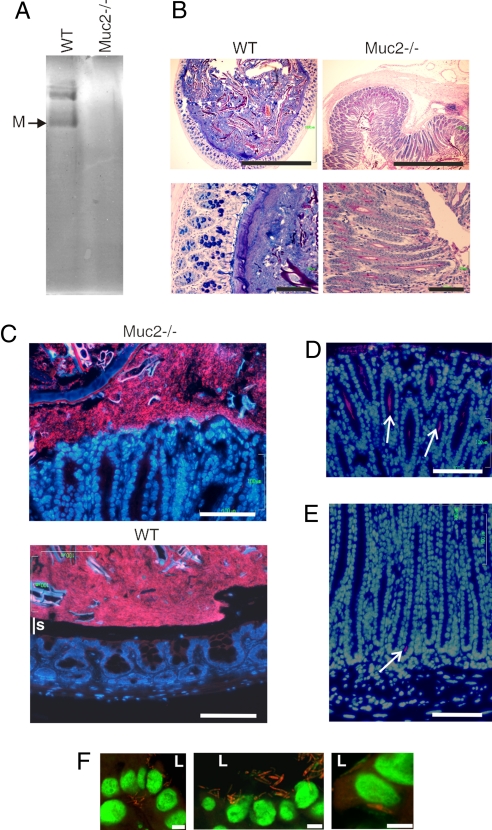
We normally live in symbiosis with approximately 10(13) bacteria present in the colon. Among the several mechanisms maintaining the bacteria/host balance, there is limited understanding of the structure, function, and properties of intestinal mucus. We now demonstrate that the mouse colonic mucus consists of two layers extending 150 mum above the epithelial cells. Proteomics revealed that both of these layers have similar protein composition, with the large gel-forming mucin Muc2 as the major structural component. The inner layer is densely packed, firmly attached to the epithelium, and devoid of bacteria. In contrast, the outer layer is movable, has an expanded volume due to proteolytic cleavages of the Muc2 mucin, and is colonized by bacteria. Muc2(-/-) mice have bacteria in direct contact with the epithelial cells and far down in the crypts, explaining the inflammation and cancer development observed in these animals. These findings show that the Muc2 mucin can build a mucus barrier that separates bacteria from the colon epithelia and suggest that defects in this mucus can cause colon inflammation.


Conflict of interest statement

The authors declare no conflict of interest.

Figures

Fig. 1.
Fig. 1.
The loose and the firm mucus layers of the colon consist mainly of the Muc2 mucin. (A) Schematic view of the domain organization of the MUC2 mucin and indication of where the sites for polymerization are localized. The human apoprotein is 5,174 aa long (2); vW identifies von Willebrand C (dark blue) or D (red) domain; PTS, the mucin domain (green); CysD (yellow), and CK, the cysteine knot domain (light blue) (1). The bands in C were digested with trypsin, and the peptides identified with LC-MS/MS and searched against the mucin database. Identified Muc2 peptides are schematically marked with one line per peptide under the Muc2 protein sequence. Peptides identified in both fractions are marked blue, those found only in the firm fraction are marked green, and those only found in the loose fraction are marked red. A full peptide list is presented in Table S1. (B) In vivo measurements of the mucus thickness in the distal colon reveal the presence of a firm and a loose layer. The thickness of the total mucus (F+L) and of the remaining firm (F) mucus after removal of the loose (L) layer is presented. The regeneration of the loose mucus is determined 15 and 30 min after its removal. C57BL/6 mice (n = 5) were analyzed, values are mean ± SEM. (C) Total loose (L) and firm (F) mucus from the measured area in B was separated on composite AgPAGE and visualized by staining the gel with Alcian blue. The fast migrating band corresponding to the smallest identified form of Muc2, most likely the monomer, is indicated by M.
Fig. 2.
Fig. 2.
The firm and loose mucus layers have different properties. (A) Loose (L) and firm (F) mucus were extracted by 6 M guanidinium chloride into soluble and insoluble pellet fractions. The insoluble pellet was photographed and is clearly visible in the firm mucus. (B) The soluble (s) and insoluble (p) fractions of loose (L) and firm (F) mucus after reduction of disulfide bonds were analyzed for mucins by AgPAGE and the gel stained with Alcian blue. The firm mucus pellet (Fp) contained Muc2 mucin, whereas the loose pellet (Lp) was almost devoid of Muc2. (C) The mucus thickness measured in rat distal colon in vivo overlaid with protease inhibitors (n = 4) or control (n = 16). The protease inhibitors (PI) were added after the first removal of the loose mucus. The firm (F) mucus is presented together with the regeneration of the loose (F+) after 20, 40, and 60 min and a final measurement of the firm (F) mucus after removal of the loose mucus. Values are mean ± SEM. *, P < 0.05 vs. untreated. The germ-free (GF) mice had a loose (white) and firm (gray) mucus layer as shown to the right (n = 5). (D) Mucus from the same measured surface area of the loose material was removed at 60 min after protease inhibitor (PI) treatment, reduced and analyzed on AgPAGE, and stained with Alcian blue. M, Muc2 monomer. (E) Western blot analysis of reduced loose and firm mucus after 60 min with or without protease inhibitor treatment. The bands were detected by the anti-MUC2N3 (anti-N3) or anti-MUC2C3 (anti-C3) antisera. (F) Equal amounts by volume of guanidinium chloride-insoluble mucus pellets from mouse colon (A) were treated or nontreated with trypsin for 3 h, resulting in volume expansion of the pellet in the trypsin-treated sample as shown by the photo and a graphic representation of the pellets.
Fig. 3.
Fig. 3.
The firm mucus layer is devoid of bacteria. (A) Muc2-positive goblet cells and overlaying mucus layers in a section of the mouse distal colon were detected by using the anti-MUC2C3 antiserum (green). The section is counterstained with DAPI to visualize nuclei. An inner stratified mucus layer (s) is linked via Muc2-stained threads (white arrow) to the goblet cells of the surface epithelia. The lowest part of the inner mucus layer is stained differently (a), suggesting a different organized form compared to the stratified (s) layer. The outer mucus layer (o) is mixed with the luminal content. (Scale bar: 50 μm.) (B) Magnification of the interface between the epithelial surface and the mucus layer, marked as in A. (C) Magnification of the inner mucus layer displayed a stratified pattern suggesting a lamellar organization. (D) Combined Muc2 immunostaining (green) as in A and FISH analysis using the general bacterial probe EUB338-Alexa Fluor 555 (red) of distal colon shows Muc2-positive goblet cells and an inner stratified (s) mucus layer on the epithelium. This layer is devoid of bacteria, which can only be detected in the outer mucus layer. The inner mucus generates a spatial separation between the cells and the microflora. (Scale bar: 20 μm.) (E) Semiquantitative PCR of bacterial 16S gene using DNA isolated from equal surface areas of the loose (L) and firm (F) mucus demonstrated more bacteria in the loose mucus layer. Amplification was done for 20, 25, and 30 cycles as indicated.
Fig. 4.
Fig. 4.
In Muc2−/− mice the bacteria are in close contact with the epithelia and enter into the crypts and cells. (A) Total mucus scraped from C57BL/6 (WT) and Muc2−/− colon were separated by reduced AgPAGE and the gel stained with Alcian blue. No major compensating mucins were observed in the sample from Muc2−/− mice as also revealed by proteomics. (B) Fixed sections of the distal colon from WT or Muc2−/− mice were stained with Alcian blue/PAS. The samples are shown in the same scale relative to each other. Muc2−/− animals have an enlarged colon diameter with elongated crypts. (Scale bars: 1 mm (Upper) and 100 μm (Lower). (C) FISH using the EUB338-Alexa Fluor 555 probe staining bacteria and DAPI DNA staining in colon show a clear separation of the bacterial DNA and epithelial surface in WT mice, but not in Muc2−/−. This separation corresponds to the stratified mucus layer (s). (Scale bar: 100 μm.) (D and E) Alcian blue/PAS and FISH staining of Muc2−/− colon show bacteria far down into the crypts as indicated by white arrows. This was never observed in WT mice. (Scale bar: 100 μm.) (F) Bacteria were detected close to the nuclei (1–5 μm) or basal of the nuclei in some epithelial and in detached cells. Confocal microscopy of a 1.5-μm optical slice with bacteria (EUB338) red and DNA green. L, lumen. (Scale bars: 5 μm.)

References

    1. Lang T, Hansson GC, Samuelsson T. Gel-forming mucins appeared early in metazoan evolution. Proc Natl Acad Sci USA. 2007;104:16209–16214. - PMC - PubMed
    1. Gum JR, Hicks JW, Toribara NW, Siddiki B, Kim YS. Molecular cloning of human intestinal mucin (MUC2) cDNA. Identification of the amino terminus and overall sequence similarity to prepro-von Willebrand factor. J Biol Chem. 1994;269:2440–2446. - PubMed
    1. Axelsson MAB, Asker N, Hansson GC. O-glycosylated MUC2 monomer and dimer from LS 174T cells are water-soluble, whereas larger MUC2 species formed early during biosynthesis are insoluble and contain nonreducible intermolecular bonds. J Biol Chem. 1998;273:18864–18870. - PubMed
    1. Godl K, et al. The N-termini of the MUC2 mucin form trimers that are held together within a trypsin-resistant core fragment. J Biol Chem. 2002;277:47248–47256. - PubMed
    1. Atuma C, Strugula V, Allen A, Holm L. The adherent gastrointestinal mucus gel layer: thickness and physical state in vivo. Am J Physiol. 2001;280:G922–G929. - PubMed

Publication types

MeSH terms