. 2008 Oct;10(10):1208-16.
doi: 10.1038/ncb1781. Epub 2008 Sep 21.

Jade-1 inhibits Wnt signalling by ubiquitylating beta-catenin and mediates Wnt pathway inhibition by pVHL

Affiliations

Jade-1 inhibits Wnt signalling by ubiquitylating beta-catenin and mediates Wnt pathway inhibition by pVHL

Vipul C Chitalia et al. Nat Cell Biol. 2008 Oct.

Abstract

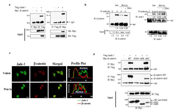
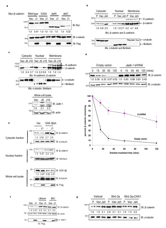
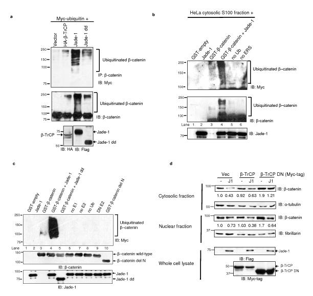
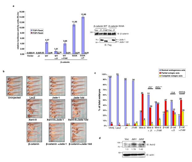
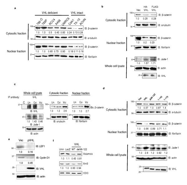
The von Hippel-Lindau protein pVHL suppresses renal tumorigenesis in part by promoting the degradation of hypoxia-inducible HIF-alpha transcription factors; additional mechanisms have been proposed. pVHL also stabilizes the plant homeodomain protein Jade-1, which is a candidate renal tumour suppressor that may correlate with renal cancer risk. Here we show that Jade-1 binds the oncoprotein beta-catenin in Wnt-responsive fashion. Moreover, Jade-1 destabilizes wild-type beta-catenin but not a cancer-causing form of beta-catenin. Whereas the well-established beta-catenin E3 ubiquitin ligase component beta-TrCP ubiquitylates only phosphorylated beta-catenin, Jade-1 ubiquitylates both phosphorylated and non-phosphorylated beta-catenin and therefore regulates canonical Wnt signalling in both Wnt-off and Wnt-on phases. Thus, the different characteristics of beta-TrCP and Jade-1 may ensure optimal Wnt pathway regulation. Furthermore, pVHL downregulates beta-catenin in a Jade-1-dependent manner and inhibits Wnt signalling, supporting a role for Jade-1 and Wnt signalling in renal tumorigenesis. The pVHL tumour suppressor and the Wnt tumorigenesis pathway are therefore directly linked through Jade-1.


Figures

Figure 1
Figure 1
Jade-1 and β-catenin interact. (a) In vivo interaction of Jade-1 and β-catenin. Extracts (600 μg protein) from transiently transfected 293T cells were immunoprecipitated (IP) with 1 μg monoclonal Myc-tag or Flag-tag antibodies. Co-immunoprecipitated β-catenin or Jade-1 was detected by immunoblotting. Whole cell lysates (WCL) (10%) were probed for input. Representative immunoblot of 4 experiments. (b) The interaction of endogenous Jade-1 and endogenous β-catenin is increased in Wnt-off phase. IPs were performed with WCL (500 μg protein) of 293T cells pretreated with vehicle (PBS + 0.1% bovine serum albumin-BSA) or 50 ng Wnt-3a ligand in PBS + 0.1% BSA, with or without 50 ng DKK1, using 1 μg of either rabbit polyclonal Jade-1 antibody (J1) or pre-immune rabbit serum (C). The co-immunoprecipitated β-catenin was detected by immunoblot with monoclonal β-catenin antibody. β-catenin was immunoprecipitated as described above using monoclonal β-catenin antibody (β-cat) and isotype control (C). Jade-1 was detected by immunoblot using Jade-1 antiserum. WCL (10%) were probed for input. Densitometry was performed to quantitate β-catenin and Jade-1. The amount of Jade-1 and β-catenin immunoprecipitated was normalized using IgG. Representative immunoblot of 3 experiments. (c) Co-localization of endogenous Jade-1 and endogenous β-catenin is increased in Wnt-off status. The 293T cells pretreated with vehicle or Wnt-3a (200 ng) for 4 h were fixed and incubated with monoclonal β-catenin and polyclonal Jade-1 antibodies followed by Alexa 594 donkey anti-mouse and Alexa 488 goat anti-rabbit as secondary antibodies. Profile plots were generated using NIH ImageJ to demonstrate quantitative Jade-1 and β-catenin protein distribution. The profile plot represents the signal intensity of each fluorophore along a single line across the midpoint of a representative cell. The X axis represents the distance in pixels through the length of a single cell, and the intensity of each fluorophore is plotted on the Y axis. A representative image from 4 experiments is shown. Scale bar = 10 μm. (d) Identification of the domain of β-catenin required for Jade-1 binding. Extracts from transiently transfected 293T cells were immunoprecipitated. WCL (10%) were probed for input. β-catenin delC and delN constructs lack the C terminus and N terminus of the protein, respectively (Supplementary Fig. S1a). β-catenin S33A is a naturally occurring, cancer-causing amino acid substitution mutant. Representative immunoblot of 3 experiments.
Figure 2
Figure 2
Jade-1 reduces β-catenin protein abundance. (a) Differential regulation of β-catenin constructs by Jade-1. Myc-tagged β-catenin and Flag-tagged Jade-1 (J1) constructs were transiently transfected into 293T cells. β-catenin construct schematics are shown in Supplementary Information, Fig. S1a. WCL were probed for β-catenin and α-tubulin protein. Expression of Jade-1 was detected by immunoblotting using Flag antibody. Densitometry was performed with NIH ImageJ using α-tubulin as loading control. Representative immunoblot of 5 experiments. (b) Jade-1 regulates the cytosolic and nuclear fractions but not the membrane fraction of endogenous β-catenin. The 293 uninfected parental cells (P) or cells infected with empty vector (Vec) or Jade-1 shRNA lentivirus (Jsh) were subjected to cell fractionation. Cytosolic, nuclear and membrane fractions were probed for β-catenin. α-tubulin, fibrillarin and E-cadherin served as loading controls and markers of cytosolic, nuclear and membrane fractions, respectively. These control blots were generated by cutting the membrane at the expected molecular weights of the controls and immunoblotting separately. One of 2 similar experiments is shown. (c) Jade-1 reduces cytosolic and nuclear pools of endogenous β-catenin. Extracts of clonal 786-O renal cancer cell lines stably expressing empty vector (Vec) or Jade-1 (J9 and J18), were probed for β-catenin. α-tubulin, fibrillarin and N-cadherin served as markers of cytosolic, nuclear and membrane fractions and as loading controls, respectively. These control blots were generated by cutting the membrane at the expected molecular weights of the controls and immunoblotting separately. Jade-1 was detected using polyclonal Jade-1 antibody. Representative immunoblot of 3 experiments. The effect of stably-transfected Jade-1 on β-catenin abundance appears to be dose dependent. (d) Jade-1 silencing substantially prolongs the half-life of endogenous cytosolic β-catenin. Digitonin was used to extract the cytosolic pool of β-catenin. Digitonin is a gently acting detergent that at low concentrations selectively punctures 8-10 nm holes in the sterol rich plasma membrane, allowing leakage of cytosolic proteins of up to 200 kDa mass (Upper panels), Digitonin extracts of 293 cells infected with empty vector or JshRNA lentiviral vector and pretreated with 20 μM emetine were probed for β-catenin by immunoblotting. (Lower panel), Percent β-catenin remaining in the digitonin-extracted fraction was analyzed by densitometry after normalizing to α-tubulin. Graph shows mean result of 4 experiments. Error bars = SEM. (e) GSK-3β silencing mitigates Jade-1 regulation of endogenous β-catenin. The 293T cells were transiently cotransfected with Flag-tagged Jade-1 (J1) and GSK-3β shRNA plasmid (GSK-3βsh) or empty vector (Vec) and GFP. One day post transfection, cells were sorted using fluorescence-activated cell sorting (FACS) and were replated for 48 hrs. Protein extracts from cells were probed for β-catenin using monoclonal β-catenin antibody. GSK-3β and Jade-1 proteins were examined by immunoblotting with polyclonal GSK-3β and Jade-1 antibodies, respectively. α-tubulin, fibrillarin and E-cadherin served as loading controls and markers of cytosolic, nuclear and membrane fractions, respectively. One of 2 similar experiments is shown. (f) Chemical inhibition of GSK-3β mitigates Jade-1 regulation of endogenous β-catenin. Transiently transfected 293T cells were serum starved for 12-16 h and treated with lithium chloride (10 mM) or BIO (100 nM) for an additional 12 h. β-catenin was detected using β-catenin antibody. Representative immunoblot of 4 experiments. (g) Jade-1 regulation of endogenous β-catenin in Wnt-on and Wnt-off status. HeLa parental cells (P) or cells infected with empty vector (Vec) or Jade-1 shRNA (Jsh) lentivirus were treated for 4 h with Wnt-3a (100 ng) with or without DKK1 (100 ng). The cytosolic fractions were probed for β-catenin and α-tubulin protein. Representative immunoblot of 4 experiments.
Figure 3
Figure 3
Jade-1 ubiquitinates β-catenin. (a) Deletion of the Jade-1 PHDs substantially reduces endogenous β-catenin ubiquitination. Extracts (600 μg protein) of 293T cells transfected and treated with MG132 (10 μM for 1 h) were immunoprecipitated with β-catenin antibody and protein A beads. The eluate was divided into two equal parts and immunoblotted separately using monoclonal Myc-tag antibody and β-catenin antibody. Expression of β-TrCP and Jade-1 protein was detected by immunoblot. One of 2 similar experiments is shown. (b) Ex vivo ubiquitination of purified β-catenin using the HeLa cell cytosolic S100 fraction. Purified recombinant GST-β-catenin on Glutathione Sepharose™ beads was incubated with HeLa cell S100 fraction (pretreated with ubiquitin aldehyde and MG132), Myc-tagged human recombinant ubiquitin (Ub), recombinant Jade-1 and energy regeneration solution (ERS). β-catenin was eluted from the beads using Laemmli buffer. The eluate was divided into two equal parts and immunoblotted separately using monoclonal Myc-tag or β-catenin antibodies. Lanes designated as no Ub or no ERS were reactions without Ub or ERS only, respectively. The eluates were also probed for Jade-1 input using polyclonal Jade-1 antibody. Lane 3 was rearranged from the same blot. β-catenin ubiquitination appears as a smear and higher molecular weight ladder. One of 2 similar experiments is shown. (c) Jade-1 ubiquitination of β-catenin in vitro, and mapping of the E3 ubiquitin ligase domain on Jade-1. A panel of E2 ubiquitin transferases, including UbcH2, UbcH3, UbcH5a, UbcH5b, UbcH5c, UbcH6, UbcH7 and UbcHc10, was screened for Jade-1-mediated β-catenin ubiquitination (data not shown). Jade-1 ubiquitinated β-catenin only in the presence of UbcH6 (Fig. 3c and Supplementary Information, Fig. S4c) or UbcH2 (data not shown). Ubiquitination reactions were reconstituted using GST-β-catenin (wild-type or delN) with E1 activating enzyme, E2 conjugase (UbcH6), E3 ligase 750 nM Jade-1 (full-length or dd), Myc-tagged human recombinant Ub and MgCl2-ATP. Lanes designated as no E1, no E2 and no Ub were reactions with all constituents except E1, E2 or Ub only, respectively. Dominant-negative UbcH6 (DN E2) was used instead of wild-type UbcH6 as a control. β-catenin was eluted from the beads using Laemmli buffer and immunoblotted using monoclonal Myc-tag antibody. GST-β-catenin and GST-Jade-1 beads equivalent to that used in the reactions were probed separately using polyclonal β-catenin C-terminal antibody and polyclonal Jade-1 antibody. Reactions without E1, E2, or ubiquitin, or with DN UbcH6, served as negative controls (lanes, 6-9). Representative immunoblot of 3 experiments. (d) Jade-1-mediated degradation of endogenous β-catenin is independent of β-TrCP. Whole cell extracts of 293T cells transfected with wild-type or DN β-TrCP, with or without Flag-tagged Jade-1, were probed using β-catenin antibody. WCL (10%) were probed using Flag- and Myc-tag antibodies for expression of Jade-1 and β-TrCP protein, respectively. α-tubulin and fibrillarin served as markers of cytosolic and nuclear fractions, respectively, and as loading controls. Representative immunoblot of 4 experiments.
Figure 4
Figure 4
Jade-1 inhibits canonical Wnt signaling. (a) Jade-1 suppresses TCF/β-catenin reporter activity. TCF-responsive promoter-reporter TOP-Flash and nonresponsive control reporter FOP-Flash were used. Activity of the Wnt signaling pathway was quantified by measuring relative firefly luciferase activity units (RLUs) normalized to renilla luciferase. The 293T cells were transfected with wild-type β-catenin (β-cat) or β-catenin S33A and Jade-1 (J1) full-length or Jade-1 dd (J1dd). Mean results of 3 experiments are shown. Error bars = SEM. Protein extracts were probed using monoclonal Flag and β-catenin antibodies to examine the expression of constructs. (b) Jade-1 inhibits Xwnt8- and β-catenin-induced ectopic axis formation in Xenopus laevis embryos. Xwnt-8 (0.34-0.45 pg) or β-catenin (100-120 pg) mRNA, with or without Jade-1 (full-length 0.8 ng or dd 1.2 ng) mRNA, was injected into ventral blastomeres. LacZ (1.5 ng) served as a negative control. A total of 7 experiments was performed to compare full-length Jade-1 and Jade-1 dd injected embryos. (c) X-wnt-8- and β-catenin-induced ectopic axis is inhibited by Jade-1 in Xenopus laevis embryos. Histogram depicting Xenopus laevis embryo phenotypes is shown. Numbers shown at the top of each bar indicate the number of embryos in each category. Chi square test was applied to determine statistical significance. *= Xwnt-8 with Jade-1 versus Xwnt-8 (p <0.0001). # = Xwnt-8 with Jade-1 dd versus Xwnt-8 (p= 0.096), **= β-catenin with Jade-1 versus β-catenin (p<0.046), §=β-catenin with Jade-1 dd versus β-catenin (p=0.087). S = statistically significant, NS = not significant. (d) Jade-1 regulates endogenous Wnt targets. WCLs of 293 cells infected with empty vector (Vec) or Jade-1 silencing lentiviral constructs (Jsh1 and Jsh2) were probed for endogenous Axin2 and actin. One of 2 similar experiments is shown.
Figure 5
Figure 5
pVHL regulates endogenous β-catenin through Jade-1. (a) β-catenin protein levels depend on VHL status in renal cancer cell lines. Cytosolic and nuclear fractions of renal-cell carcinoma cell lines were probed for endogenous β-catenin protein. One of 2 similar experiments is shown. (b) pVHL down-regulates endogenous β-catenin. Extracts from 786-O cells stably expressing empty vector (Vec) or pVHL were probed for endogenous β-catenin protein. Jade-1 and pVHL expression was detected using polyclonal Jade-1 and monoclonal pVHL antibodies. Representative immunoblot of 3 experiments. (c) pVHL knock-down increases endogenous β-catenin abundance. To confirm pVHL knock-down, pVHL was immunoprecipitated using polyclonal pVHL or control (C) antibody from untransfected (Un) 293T cells or 293T cells transfected with VHL (VHLsi) or control siRNA (Csi) oligonucleotides because endogenous pVHL abundance is low. Cell fractions were probed for endogenous Jade-1 and endogenous β-catenin protein. Representative immunoblot of 3 experiments. (d) Differential regulation of endogenous β-catenin by wild-type pVHL and pVHL truncations that do not stabilize Jade-1. Extracts of 293T cells transfected with wild-type pVHL, pVHL del96-122 (Δ96-122), pVHL 1-143 and pVHL 1-175 were probed for pVHL, endogenous β-catenin and endogenous Jade-1 by immunoblot. One of 2 similar experiments is shown. (e) pVHL suppresses endogenous Wnt targets. WCLs of empty vector (Vec) or pVHL-expressing 786-O cell lines were probed for pVHL and endogenous LEF1 and cyclin D1. Representative immunoblot of 3 experiments. (f) Wild-type pVHL, but not pVHL del96-122, suppresses β-catenin/Tcf target genes. In vitro transcribed, capped LacZ (0.288 ng), wild-type VHL (WT) (0.194 ng) or VHL del96-122 (0.293 ng) mRNA was injected into dorsal blastomeres of Xenopus laevis embryos. Injected embryos were harvested at stage 10 for RT-PCR. Semiquantitative RT-PCR analysis of the Wnt targets Xsiamois and Xnr3 was performed in whole embryos. ODC served as control. Unin.= uninjected.

Comment in

References

    1. Maxwell PH, et al. The tumour suppressor protein VHL targets hypoxia-inducible factors for oxygen-dependent proteolysis. Nature. 1999;399:271–5. - PubMed
    1. Cohen HT, McGovern FJ. Renal-cell carcinoma. N Engl J Med. 2005;353:2477–90. - PubMed
    1. Zhou MI, et al. The von Hippel-Lindau tumor suppressor stabilizes novel plant homeodomain protein Jade-1. J Biol Chem. 2002;277:39887–98. - PubMed
    1. Zhou MI, Wang H, Foy RL, Ross JJ, Cohen HT. Tumor suppressor von Hippel-Lindau (VHL) stabilization of Jade-1 protein occurs through plant homeodomains and is VHL mutation dependent. Cancer Res. 2004;64:1278–86. - PubMed
    1. Zhou MI, et al. Jade-1, a candidate renal tumor suppressor that promotes apoptosis. Proc Natl Acad Sci U S A. 2005;102:11035–40. - PMC - PubMed

Publication types