

doi: 10.1038/nmeth.1253.
Epub 2008 Sep 21.
Imaging individual mRNA molecules using multiple singly labeled probes
Affiliations
- PMID: 18806792
- PMCID: PMC3126653
- DOI: 10.1038/nmeth.1253
Item in Clipboard
Imaging individual mRNA molecules using multiple singly labeled probes
Nat Methods.
2008 Oct.
Abstract
We describe a method for imaging individual mRNA molecules in fixed cells by probing each mRNA species with 48 or more short, singly labeled oligonucleotide probes. This makes each mRNA molecule visible as a computationally identifiable fluorescent spot by fluorescence microscopy. We demonstrate simultaneous detection of three mRNA species in single cells and mRNA detection in yeast, nematodes, fruit fly wing discs, and mammalian cell lines and neurons.
Figures
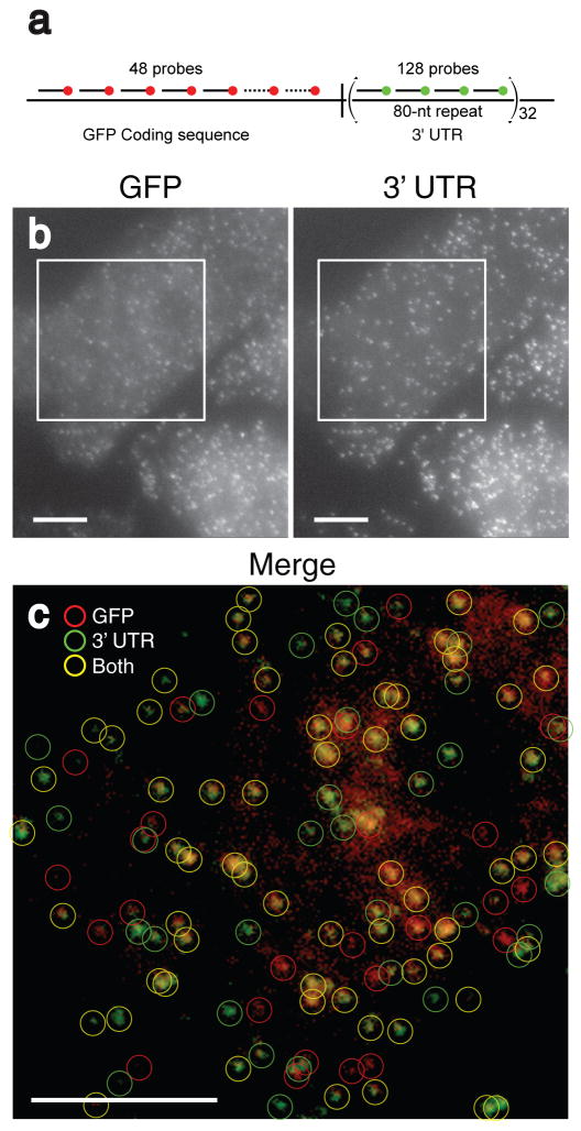
Simultaneous detection of a unique sequence and a repeated sequence in individual mRNA molecules. a) Schematic depiction of the construct used. The 48 probes used to detect the GFP coding sequence were labeled with Alexa 594 and the four different probes used to detect the tandem repeat in the 3′ UTR were labeled with TMR. b) Maximum intensity merges of a pair of z-stacks of fluorescent images of CHO cells taken in the Alexa 594 channel (left) and the TMR channel (right) corresponding to GFP coding region probes and UTR probes, respectively. c) False color merge of the images in b) enclosed by the squares, with circles representing computationally identified mRNA particles. All scale bars are 5 μm long.
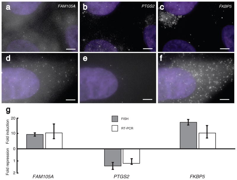
Simultaneous imaging of three different mRNAs in mammalian cells. a–c) Images showing FAM105A, PTGS2 and FKBP5 mRNA particles in the same set of A549 cells not treated with dexamethasone. d–f) Images showing FAM105A, PTGS2 and FKBP5 particles in cells treated for 8 hours with 24 nM dexamethasone. g) Fold induction for all three genes as measured by FISH and real-time RT-PCR; error bars for FISH were obtained by bootstrapping and those for RT-PCR were obtained by repetition as described in the supplementary information. All images are maximum merges of a z-stack of fluorescent images spanning the extent of the cells with nuclear DAPI counterstaining in purple, and all scale bars are 5 μm long.
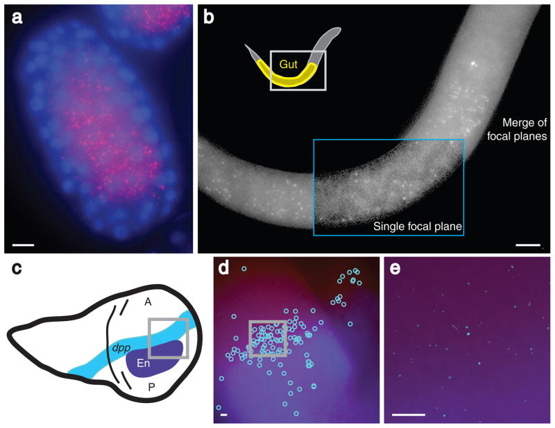
Imaging localized mRNAs in C. elegans and D. melanogaster. a) elt-2 mRNA molecules (red) in an early stage embryo (~100 cell stage) from C. elegans; the nuclei have been counterstained with DAPI (blue). b) elt-2 mRNA molecules in an L1 larva from C. elegans. Inside the blue box, a single focal plane is shown in which the intestinal track is visible. c) A schematic depiction of dpp and engrailed expression in the imaginal wing discs of third instar larvae from D. melanogaster. d) Image showing the locations of the computationally identified dpp mRNA molecules (light blue circles) and Engrailed expression detected by immunofluorescence (dark blue). e) Image containing enhanced dpp mRNA molecule signals (light blue) and Engrailed protein expression detected by immunofluorescence (dark blue). All images except the boxed portion of (b) are maximum merges of a z-stack of fluorescent images, and all scale bars are 5 μm long.
References
-
- Kaufmann BB, van Oudenaarden A. Stochastic gene expression: from single molecules to the proteome. Curr Opin Genet Dev. 2007;17:107–112. - PubMed
-
- St Johnston D. Moving messages: the intracellular localization of mRNAs. Nat Rev Mol Cell Biol. 2005;6:363–375. - PubMed
-
- Levsky JM, Singer RH. Fluorescence in situ hybridization: past, present and future. J Cell Sci. 2003;116:2833–2838. - PubMed
-
- Raap AK, et al. Ultra-sensitive FISH using peroxidase-mediated deposition of biotin- or fluorochrome tyramides. Hum Mol Genet. 1995;4:529–534. - PubMed
Publication types
MeSH terms
Substances
Grants and funding
LinkOut - more resources
Full Text Sources
Other Literature Sources
Molecular Biology Databases
