. 2008 Sep 22;3(9):e3256.
doi: 10.1371/journal.pone.0003256.

Induction of pluripotent protective immunity following immunisation with a chimeric vaccine against human cytomegalovirus

Affiliations

Induction of pluripotent protective immunity following immunisation with a chimeric vaccine against human cytomegalovirus

Jie Zhong et al. PLoS One. .

Abstract

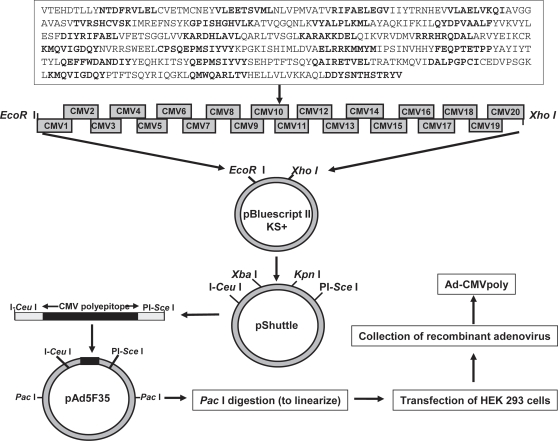
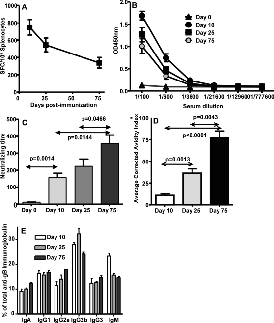
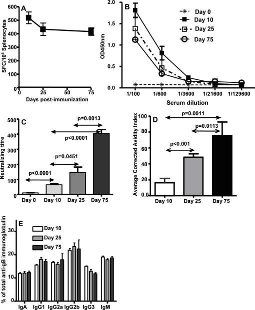
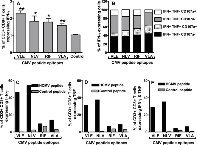
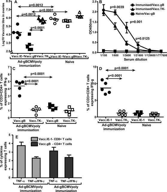
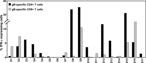
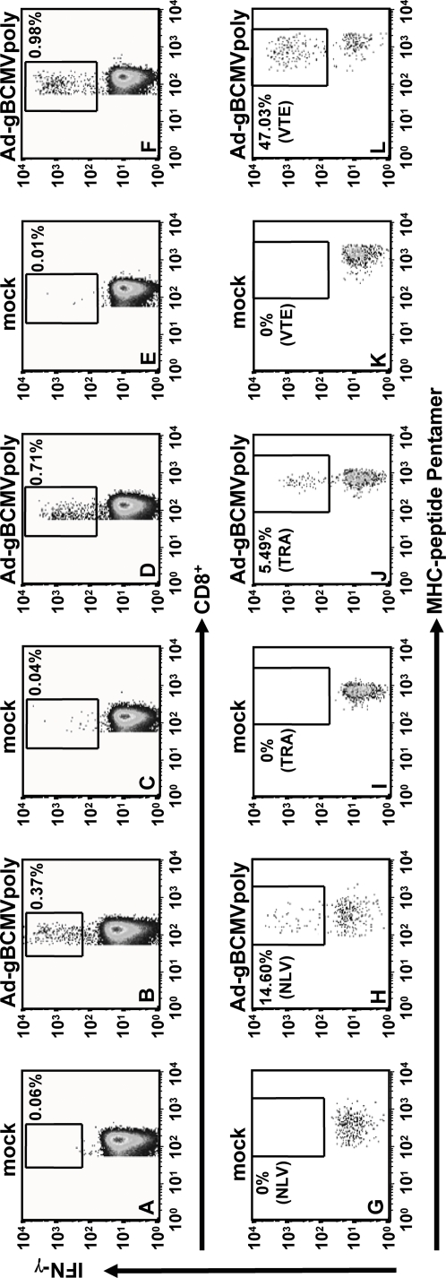
Based on the life-time cost to the health care system, the Institute of Medicine has assigned the highest priority for a vaccine to control human cytomegalovirus (HCMV) disease in transplant patients and new born babies. In spite of numerous attempts successful licensure of a HCMV vaccine formulation remains elusive. Here we have developed a novel chimeric vaccine strategy based on a replication-deficient adenovirus which encodes the extracellular domain of gB protein and multiple HLA class I & II-restricted CTL epitopes from HCMV as a contiguous polypeptide. Immunisation with this chimeric vaccine consistently generated strong HCMV-specific CD8(+) and CD4(+) T-cells which co-expressed IFN-gamma and TNF-alpha, while the humoral response induced by this vaccine showed strong virus neutralizing capacity. More importantly, immunization with adenoviral chimeric vaccine also afforded protection against challenge with recombinant vaccinia virus encoding HCMV antigens and this protection was associated with the induction of a pluripotent antigen-specific cellular and antibody response. Furthermore, in vitro stimulation with this adenoviral chimeric vaccine rapidly expanded multiple antigen-specific human CD8(+) and CD4(+) T-cells from healthy virus carriers. These studies demonstrate that the adenovirus chimeric HCMV vaccine provides an excellent platform for reconstituting protective immunity to prevent HCMV diseases in different clinical settings.


Conflict of interest statement

Competing Interests: The authors have declared that no competing interests exist.

Figures

Figure 1
Figure 1. Schematic representation of the construction of a recombinant adenovirus that expresses a synthetic DNA encoding for a polyepitope protein which contains 46 HCMV T-cell epitopes (see box and Table 1).
Each of the alternate epitope sequences are shown in bold letters. The DNA sequence encoding this polyepitope protein was constructed using overlapping epitope sequence specific primers (referred to as CMV1 to CMV20) as described in the “Material and Methods” section. This synthetic insert was first cloned into a pBluescript II KS+ phagemid, prior to cloning into the pShuttle vector. After amplification in E.coli, the expression cassette from pShuttle was excised and ligated into the Ad5F35 expression vector. Following linearization of the DNA using Pac I restriction enzyme, the recombinant Ad5F35 vector was packaged into infectious adenovirus by transfecting HEK 293 cells, and recombinant adenovirus (referred to as Ad-CMVpoly) was harvested from transfected cells by repeated freeze-thawing cycles.
Figure 2
Figure 2. HCMV epitope-specific T cell response following primary and secondary immunisation with Ad-CMVpoly vaccine.
Two different groups of HHD-2 mice were immunised intramuscularly with Ad-CMVpoly (7.5×10⁸ PFU/mouse). A & B, Following primary immunisation, animals were sacrificed 10 days post immunisation and HCMV epitope-specific reactivity was assessed in the splenocytes by ELISPOT assays as described in the “Material and Methods” section. The epitopes tested for T cell reactivity were VLE (IE-1), NLV (pp65), RIF (pp65), VLA (IE-1), IIY (IE-2) AVG (gB). C & D, For immunological analysis following secondary immunisation, animals were given booster immunisation (7.5×10⁸ PFU/mouse) 100 days after primary immunisation and then sacrificed 10 days post secondary immunisation. HCMV epitope-specific reactivity was assessed as described above. A & C shows ELISPOT data based on the pooled HLA A2-restricted HCMV epitopes, while B & D shows relative T cell responses to individual epitopes. The results are expressed as Mean±SE of spot forming cells (SFC) per 106 splenocytes from four individually tested mice. E, Anti-adenovirus antibody titre induced by immunisation with Ad-CMVpoly. Serum samples were collected at different time points after immunisation and anti-adenovirus titres were evaluated by ELISA as described in the “Material and Methods” section. All statistical analyses were conducted using GraphPad Prism 4 software.
Figure 3
Figure 3. HCMV-specific effector and memory cellular and humoral immune responses following immunisation with a mixture of Ad-CMVpoly and Ad-gB vaccines.
A, HCMV-specific CD8+ T cell responses following immunisation with Ad-CMVpoly and Ad-gB. These T cell responses were assessed using ELISPOT assays on day 10, 25 and 75 post immunisation. The results are expressed as Mean±SE of spot forming cells (SFC) per 106 splenocytes. B, gB-specific antibody responses in serum samples from immunised mice on days 10, 25 and 75. Serum samples on day 0 were collected before the immunisation. C, Virus neutralizing capacity of antibody responses induced in HHD-2 mice immunised with Ad-CMVpoly and Ad-gB. Serum samples from these mice were pre-incubated with HCMV virus Ad169 and then these virus preps were used to infect MRC-5. Following overnight incubation virus infectivity was assessed using IE-1/IE-2 expression as outlined in the “Material and Methods” section. D, Avidity maturation of gB-specific antibody responses in Ad-CMVpoly and Ad-gB immunised mice. E, Immunoglobulin subclass analysis of gB-specific antibody responses in HHD-2 vaccinated mice. Serum samples were collected from three different groups of mice on days 10, 25 and 75 post-immunisation. A minimum of five mice from each group were assessed for HCMV epitope-specific T cell reactivity and humoral immune responses. All statistical analyses were conducted using GraphPad Prism 4 software.
Figure 4
Figure 4. HCMV-specific effector and memory humoral and cellular immune responses following immunisation with Ad-gBCMVpoly vaccine.
A, HCMV-specific CD8+ T cell responses following immunisation with Ad-gBCMVpoly. These T cell responses were assessed using ELISPOT assays on day 10, 25 and 75 post immunisation. The results are expressed as Mean±SE of spot forming cells (SFC) per 106 splenocytes. B, gB-specific antibody responses in serum samples from immunised mice on days 10, 25 and 75. Serum samples on day 0 were collected before the immunisation. C, Virus neutralizing capacity of antibody responses induced following immunisation with Ad-gBCMVpoly. Serum samples from these mice were pre-incubated with HCMV virus Ad169 and then these virus preps were used to infect MRC-5. Following overnight incubation virus infectivity was assessed using IE-1/IE-2 expression as outlined in the “Material and Methods” section. D, Avidity maturation of gB-specific antibody responses in Ad-gBCMVpoly immunised mice. E, Immunoglobulin subclass analysis of gB-specific antibody responses in HHD-2 vaccinated mice. Serum samples were collected from three different groups of mice on days 10, 25 and 75 post-immunisation. A minimum of five mice from each group were assessed for HCMV epitope-specific T cell reactivity and humoral immune responses. All statistical analyses were conducted using GraphPad Prism 4 software.
Figure 5
Figure 5. Cytokine expression by HCMV-specific CD8+ T cells from Ad-gBCMVpoly immunised HHD-2 mice.
A & B, Ex vivo expression of IFN-γ, TNF-α and CD107a by antigen-specific CD8+ T-cells from Ad-gBCMVPpoly vaccinated mice 10 days post-vaccination. Splenocytes were prepared from 3 individual HHD-2 mice 10 days post-vaccination and cultured with individual HCMV peptides overnight. Anti-CD107a antibody and Brefeldin A was added during the last 6 and 5 hours incubation respectively, followed by T cell surface marker and intracellular cytokine staining. Data represent the percentage of IFN-γ expressing CD8+ T cells (A) and percentage of single, double or triple markers expressing cells among IFN-γ expressing CD8+ T cells (B). C–E, Expression of IFN-γ and/or TNF-α by in vitro expanded antigen-specific CD8+ T-cells from Ad-gBCMVPpoly vaccinated mice 10 days post-vaccination. Splenocytes pooled from three immunised mice were first stimulated with individual HCMV peptide epitope-pulsed splenocytes for 2 weeks in the presence of recombinant mouse IL-2 at the concentration of 10 IU/ml, then cultured with MRC-5 cells pulsed with corresponding HCMV peptide epitope overnight for intracellular cytokine assay. The HCMV peptide epitopes tested here were VLE (IE-1), NLV (pp65), RIF (pp65), VLA (IE-1) at the concentration of 1 µg/ml. Data represent the percentage of IFN-γ (C), TNF-α (D) and IFN-γ & TNF-α (E) expressing CD8+ T-cells. ** (p<0.005) and * (p<0.05) show statistically significant difference between indicated CMV peptide epitopes and control epitope (A). Data from one out three experiments with similar results was shown in C–E. All statistical analyses were conducted using GraphPad Prism 4 software.
Figure 6
Figure 6. Ad-gBCMVpoly induced protection against challenge with recombinant vaccinia expressing gB or IE-1 protein.
HHD-2 mice were immunised with Ad-gbCMVpoly vaccine and 21 days following vaccination these mice were challenged (intraperitoneal) with recombinant vaccinia encoding gB (Vacc.gB), IE1 protein (Vacc.IE-1) or control vaccinia (Vacc.TK) at 107 pfu virus/mouse. Ovaries, splenocytes and peripheral blood samples were collected four days later and used for assessing viral load, antigen-specific T cell response and gB-specific antibody response. A, Virus titres in the ovaries of Ad-gBCMVpoly immunised or naïve HHD-2 mice challenged with Vacc.IE-1, Vacc.gB or Vacc.TK. B, gB-specific antibody response in Ad-gBCMVpoly immunised or naïve HHD-2 mice challenged with Vacc.gB or Vacc.TK. C, Ex vivo gB-specific CD3+CD4+ T cell response in Ad-gBCMVpoly immunised or naïve HHD-2 mice challenged with Vacc.gB or Vacc.TK. Splenocytes from these mice were stimulated with recombinant gB protein (40 µg/ml) overnight and then assessed for IFN-γ production using intracellular cytokine assay. D, Ex vivo IE-1-specific CD3+CD8+ T cell response in Ad-gBCMVpoly immunised or naïve HHD-2 mice challenged with Vacc.IE-1 or Vacc.TK. Splenocytes from these mice were stimulated with the peptide epitope VLEETSVML (1 µg/ml) overnight and then assessed for IFN-γ production using intracellular cytokine assay. E, Ex vivo expression of IFN-γ and/or TNF-α by antigen-specific CD8+ and CD4+T-cells from Ad-gBCMVPpoly vaccinated mice, challenged with recombinant vaccinia encoding IE-1 or gB. Splenocytes from immunised mice stimulated with either gB protein or IE-1 peptide epitope overnight for intracellular cytokine assay. Data represent the percentage of TNF-α and IFN-γ & TNF-α expressing CD4+ or CD8+ T-cells. All statistical analyses were conducted using GraphPad Prism 4 software.
Figure 7
Figure 7. Expansion of gB-specific T cells following in vitro stimulation of human PBMC with Ad-gBCMVpoly.
PBMC from a panel of healthy virus carriers (referred to as D1–D17) were co-cultured with autologous PBMC infected with Ad-gBCMVpoly (MOI: 5∶1 or 1∶1) at a responder to stimulator ratio of 2∶1. These cultures were supplemented with rIL-2 (10 U/ml) on day 3 and every 3–4 days thereafter. On day 14, these T cell cultures were tested against a panel of pooled overlapping gB peptides (20 aa long, overlapping by 10 aa) using intracellular cytokine assays. The data presented in the figure shows the percentage of gB-specific CD8+ and CD4+ T cell recovered from each donor following stimulation with Ad-gBCMVpoly.
Figure 8
Figure 8. Ex vivo stimulation of human PBMC with Ad-gBCMVpoly.
PBMC from healthy virus carriers were co-cultured with autologous PBMC infected with Ad-gBCMVpoly (MOI: 5∶1) at a responder to stimulator ratio of 2∶1 for 6 h. These T cells were then co-stained with anti-CD3, anti-CD8, PE-labelled anti-INF-γ antibody and APC-labelled MHC-peptide multimers. A–F, Percentage of CD8+ T cells expressing INF-γ following mock stimulation or Ad-gBCMVpoly stimulation. G–L, Percentage of MHC-peptide pentamer-positive cells expressing IFN-γ following mock stimulation or Ad-gBCMVpoly stimulation. Pentamers used for each of the HCMV epitopes are indicated in G–L.

Similar articles

Cited by

References

    1. Gandhi MK, Khanna R. Human cytomegalovirus: clinical aspects, immune regulation, and emerging treatments. Lancet Infect Dis. 2004;4:725–738. - PubMed
    1. Gaytant MA, Steegers EA, Semmekrot BA, Merkus HM, Galama JM. Congenital cytomegalovirus infection: review of the epidemiology and outcome. Obstet Gynecol Surv. 2002;57:245–256. - PubMed
    1. Arvin AM, Fast P, Myers M, Plotkin S, Rabinovich R. Vaccine development to prevent cytomegalovirus disease: report from the National Vaccine Advisory Committee. Clin Infect Dis. 2004;39:233–239. - PubMed
    1. Stratton KR, Durch JS, Lawrence RS Committee to Study Priorities for Vaccine D. Vaccines for the 21st Century: A tool for decision making. Bethesda: National Academy Press; 2001. p. 476.
    1. Reddehase MJ. Antigens and immunoevasins: opponents in cytomegalovirus immune surveillance. Nat Rev Immunol. 2002 Nov;2(11):831–844. - PubMed

Publication types