. 2008;4(1-2):33-52.
doi: 10.1007/s10404-007-0198-8.

Nanobiosensors: optofluidic, electrical and mechanical approaches to biomolecular detection at the nanoscale

Affiliations

Nanobiosensors: optofluidic, electrical and mechanical approaches to biomolecular detection at the nanoscale

David Erickson et al. Microfluid Nanofluidics. 2008.

Abstract

Next generation biosensor platforms will require significant improvements in sensitivity, specificity and parallelity in order to meet the future needs of a variety of fields ranging from in vitro medical diagnostics, pharmaceutical discovery and pathogen detection. Nano-biosensors, which exploit some fundamental nanoscopic effect in order to detect a specific biomolecular interaction, have now been developed to a point where it is possible to determine in what cases their inherent advantages over traditional techniques (such as nucleic acid microarrays) more than offset the added complexity and cost involved constructing and assembling the devices. In this paper we will review the state of the art in nanoscale biosensor technologies, focusing primarily on optofluidic type devices but also covering those which exploit fundamental mechanical and electrical transduction mechanisms. A detailed overview of next generation requirements is presented yielding a series of metrics (namely limit of detection, multiplexibility, measurement limitations, and ease of fabrication/assembly) against which the various technologies are evaluated. Concluding remarks regarding the likely technological impact of some of the promising technologies are also provided.


Figures

Fig. 1
Fig. 1
Four-channel Young’s interferometer based optical biosensor from Ymeti et al. (2007). Channels 1, 2, and 3 are the sensor channels which are functionalized with antibody capture probes and 4 is the reference channel. Copyright American Chemical Society. Reproduced with permission
Fig. 2
Fig. 2
Optically resonant photonic crystal biosensor from Lee and Fauchet (2007). Scanning electron microscopy photograph of a typical device used in these experiments and schematic of the experimental setup. A limit of detection (LOD) on the order of 2.5 fg (for bovine serum albumin) was obtained using this device. Copyright Optical Society of America. Reproduced with permission
Fig. 3
Fig. 3
Variations on surface plasmon resonance (SPR) detection. a Angular SPR, b spectral SPR, and c nanoparticle or Local SPR. Details on each of the above techniques are provided in the text
Fig. 4
Fig. 4
Dark-field optical image of Ag nanoparticles used as single particle nanosensors in McFarland and Van Duyne (2003). The field of view in this image is approximately 130 µm × 170 µm. Copyright Optical Society of America. Reproduced with permission
Fig. 5
Fig. 5
Surface plasmon resonance antibody array chip from Usui-Aoki et al. (2005). Antibody microarray layout. SPR signals from this array were measured by a FLEXCHIP™ Kinetic Analysis System. a Layout of the array containing 400 reaction sites which could be monitored in parallel. Array details are available in the aforementioned reference. b Overview of the affinity chip where all reaction sites are located in a 1 cm × 1 cm. A flow cell with a volume of 47 µL is used to transport the sample over the array. c Visualization of immobilized antibodies. Copyright Wiley-VCH Verlag GmbH & Co KGaA. Reproduced with permission
Fig. 6
Fig. 6
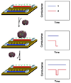
Technique for semiconductor nanowire based detection of single viruses from Patolsky et al. (2004). In this figure, two nanowire devices (labeled 1 and 2) are functionalized with different antibodies. The antibodies on the second wire are specific to the target virus. When introduced, the virus binds to the second nanowire and a change in the conductance is observed and taken to be indicative of the presence of the virus in solution. When the virus unbinds from the nanowire, the conductance returns to its original value. Figure is Copyright 2004 National Academy of Sciences, USA
Fig. 7
Fig. 7
Nanomechanical resonators for ultralow LOD mass sensors from Ilic et al. (2004a, b). Oblique-angle SEM micrographs of ad cantilevers (scale bar = 5 µm) and eh bridge oscillators (scale bar = 2 µm). The diameters of the Au pads were 50, 100, 200, and 400 nm, from left to right. Reused with permission from (Ilic et al. 2004a, b). Copyright 2004, American Institute of Physics

References

    1. Armani AM, Vahala KJ. Heavy water detection using ultrahigh-Q microcavities. Opt Lett. 2006;31(12):1896–1898. - PubMed
    1. Armani DK, Kippenberg TJ, Spillane SM, Vahala KJ. Ultrahigh-Q toroid microcavity on a chip. Nature. 2003;421(6926):925–928. - PubMed
    1. Arnold S, Khoshsima M, Teraoka I, Holler S, Vollmer F. Shift of whispering-gallery modes in microspheres by protein adsorption. Opt Lett. 2003;28(4):272–274. - PubMed
    1. Aslan K, Lakowicz JR, Geddes CD. Nanogold-plasmon-resonance-based glucose sensing. Anal Biochem. 2004;330(1):145–155. - PMC - PubMed
    1. Bard A. Electrochemical methods: fundamentals and applications. New York: Wiley; 2001.

LinkOut - more resources