. 2008 Oct 15;130(41):13696-708.
doi: 10.1021/ja803928m. Epub 2008 Sep 23.

Testing geometrical discrimination within an enzyme active site: constrained hydrogen bonding in the ketosteroid isomerase oxyanion hole

Affiliations

Testing geometrical discrimination within an enzyme active site: constrained hydrogen bonding in the ketosteroid isomerase oxyanion hole

Paul A Sigala et al. J Am Chem Soc. .

Abstract

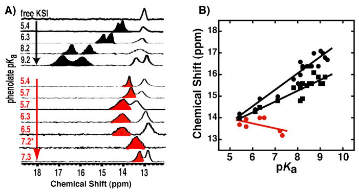
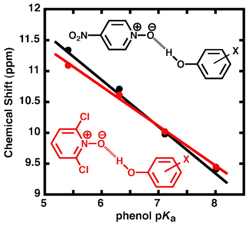
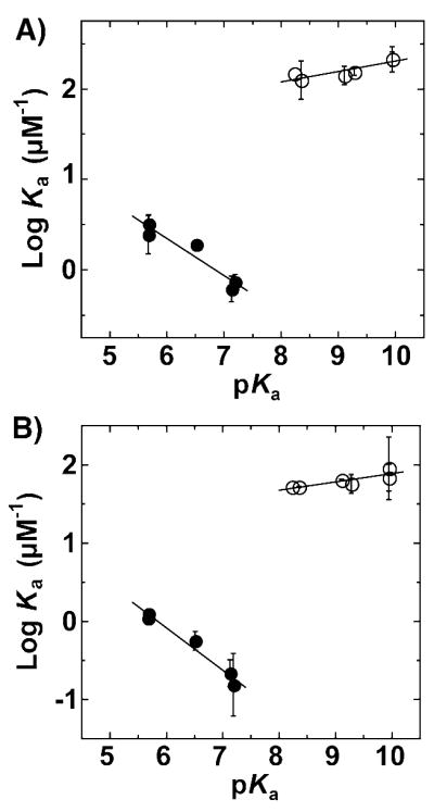
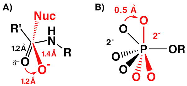
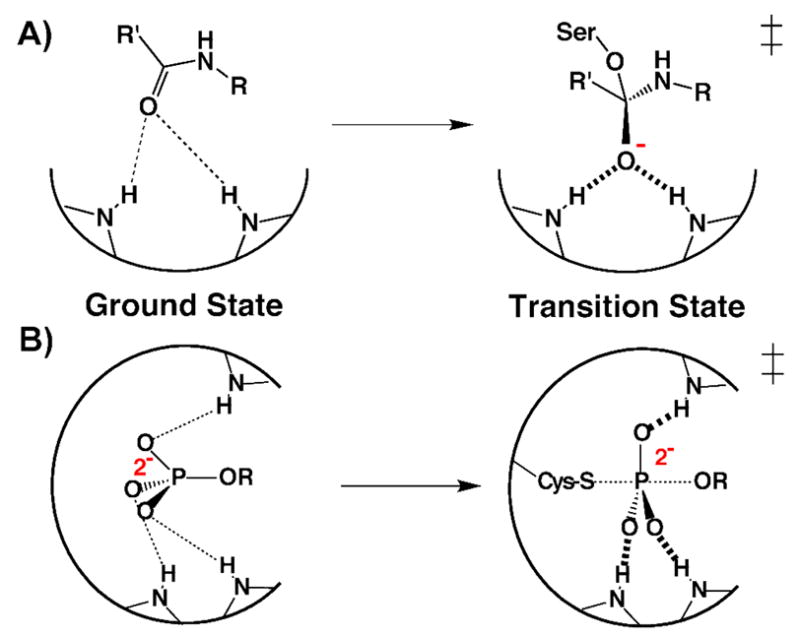
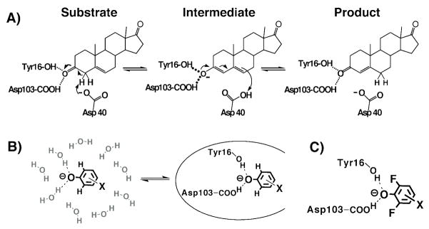
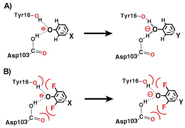
Enzymes are classically proposed to accelerate reactions by binding substrates within active-site environments that are structurally preorganized to optimize binding interactions with reaction transition states rather than ground states. This is a remarkably formidable task considering the limited 0.1-1 A scale of most substrate rearrangements. The flexibility of active-site functional groups along the coordinate of substrate rearrangement, the distance scale on which enzymes can distinguish structural rearrangement, and the energetic significance of discrimination on that scale remain open questions that are fundamental to a basic physical understanding of enzyme active sites and catalysis. We bring together 1.2-1.5 A resolution X-ray crystallography, (1)H and (19)F NMR spectroscopy, quantum mechanical calculations, and transition-state analogue binding measurements to test the distance scale on which noncovalent forces can constrain the structural relaxation or translation of side chains and ligands along a specific coordinate and the energetic consequences of such geometric constraints within the active site of bacterial ketosteroid isomerase (KSI). Our results strongly suggest that packing and binding interactions within the KSI active site can constrain local side-chain reorientation and prevent hydrogen bond shortening by 0.1 A or less. Further, this constraint has substantial energetic effects on ligand binding and stabilization of negative charge within the oxyanion hole. These results provide evidence that subtle geometric effects, indistinguishable in most X-ray crystallographic structures, can have significant energetic consequences and highlight the importance of using synergistic experimental approaches to dissect enzyme function.


Figures

Figure 1
Figure 1
Crystal structure of DFP bound at the active site of pKSID40N. (A) Sigma-A weighted 2Fo – Fc electron density map (contoured at 1.3 σ) showing DFP (fluorine atoms in purple) bound in the oxyanion hole, positioned to receive short hydrogen bonds from Tyr16 and Asp103. (B) Superposition of the pKSID40N•DFP (carbon atoms in yellow) and pKSID40N•phenolate structures (carbon atoms in green), highlighting the short O•••O and O•••F distances to DFP. (C) Side-view of the overlay in (B), looking down the phenolate C-O axis and highlighting the different angular displacements for hydrogen bonds formed to the two ligands above and below the plane of the phenolate ring.
Figure 2
Figure 2
Structural features of KSI-bound DFP and the surrounding binding pocket. (A) Van der Waals protein surface (blue) in the oxyanion hole, showing the close packing of groups around the bound DFP (van der Waals surface shown as dots and colored by atom type: yellow, carbon; red, oxygen; purple, fluorine) and side chains of Asp103 and Tyr16 (yellow). (B) Energy minimized gas-phase structure (blue) of DFP receiving hydrogen bonds from Y16 (4-Me-phenol) and D103 (acetic acid), calculated at the B3LYP level using the 6-311++G(d,p) basis set, overlaid with the corresponding side chain and DFP orientations observed in the pKSID40N•DFP X-ray structure (yellow). (C) pKSID40N backbone colored according to the highest atomic B factor observed for each residue. The Asp103 and Tyr16 side chains are shown color-coded by their atomic B-factors. The DFP ligand is in yellow (not color-coded by B-factor).
Figure 3
Figure 3
19F NMR spectra of tKSID40N•DFP complexes. (A) 19F NMR chemical shifts of the meta- and para- F atoms in KSI-bound 3,4,5-trifluorophenolate (KSI•RO) are nearly identical to those of the free phenolate (RO-) in solution at pH 12 (from ref. 55). (B) 19F NMR chemical shifts of the ortho-F atoms in KSI-bound 3-methyl-2,6-difluorophenolate are perturbed downfield from their values when free in solution.
Figure 4
Figure 4
(A) Superposition of the pKSID40N•2-F-phenolate (orange) and pKSID40N•phenolate (green) structures. (B) Side-view of the overlay in (A), looking down the phenolate C-O axis. (C) 19F NMR spectra of 2-F-4-NO2-phenolate free in solution at pH 12 (RO) and bound to tKSID40N (KSI•RO).
Figure 5
Figure 5
1H NMR spectra of tKSID40N•phenolate complexes. (A) Downfield regions of spectra of non-ortho-substituted phenolates (black, from ref. 55), and DFPs (red) bound to KSI at pH 7.2. Note the spectrum for 2,4,6-F3-phenol (pKa 7.2) was acquired at pH 5.8 due to substantial overlap of the hydrogen-bonded peak with the 13 ppm enzymatic peak at pH 7.2 (see Figure S4). (B) Correlation between increasing phenolate pKa and chemical shift of the observed downfield hydrogen bonded proton peak(s). Black symbols represent the two black peaks observed in the non-ortho-substituted phenolate spectra in (A) (and additional spectra not shown; data from ref. 55), and red symbols represent the downfield peak observed in the DFP spectra in (A) (data from Table S3).
Figure 6
Figure 6
Dependence of the hydrogen bonded proton chemical shift on phenol pKa for a series of phenol donors (3,4-dinitrophenol, 3-CF3-4-nitrophenol, 4-nitrophenol, and 4-cyanophenol) hydrogen-bonded to either 4-nitropyridine-N-oxide (black, pKa = −1.7) or 2,6-dichloropyridine-N-oxide (red, pKa = − 2.3) in chloroform (data from Table S4).
Figure 7
Figure 7
Energetics of fluorophenolate binding to KSID40N. The dependence of affinity (log Ka) on the pKa of non-ortho-fluorophenolates (open circles; from ref. 55) and di-ortho-fluorophenolates (filled circles) for binding to pKSI (A) and tKSI (B). Slopes for pKSI are 0.11 ± 0.03 and −0.41 ± 0.08 and for tKSI are 0.10 ± 0.03 and −0.54 ± 0.06. (Data from Table S5.)
Scheme 1
Scheme 1
Geometric and electrostatic changes in the (A) carbonyl C-O during peptide bond hydrolysis and (B) nonbridging oxygen atoms during phosphoryl transfer reactions (nucleophile not shown) going from the ground state (black) to the transition state (red).
Scheme 2
Scheme 2
Schematic depiction of proposed optimization of transition state rather than ground state binding within the (A) serine protease and (B) protein tyrosine phosphatase active sites.
Scheme 3
Scheme 3
(A)Mechanism of KSI catalyzed steroid isomerization. (B) Equilibrium binding of a substituted phenolate from water into the KSI active site. (C) Schematic depiction of a di-ortho-F-substituted phenolate bound at the KSI active site.
Scheme 4
Scheme 4
Physical model for phenolate binding in the KSI oxyanion hole. (A) Hydrogen bonds formed to meta-and para-substituted phenolates shorten with increasing phenolate charge localization (indicated by larger minus sign). (B) O•••F contacts constrain hydrogen bonds formed to DFPs from shortening with increasing phenolate charge localization.

References

    1. Lad C, Williams NH, Wolfenden R. Proc Natl Acad Sci. 2003;100:5607–5610. - PMC - PubMed
    1. Bolon DN, Voigt CA, Mayo SL. Curr Opin Chem Biol. 2002;6:125–129. - PubMed
    1. Jiang L, Althoff EA, Clemente FR, Doyle L, Rothlisberger D, Zanghellini A, Gallaher JL, Betker JL, Tanaka F, Barbas CF, 3rd, Hilvert D, Houk KN, Stoddard BL, Baker D. Science. 2008;319:1387–1391. - PMC - PubMed
    1. Kaplan J, DeGrado WF. Proc Natl Acad Sci. 2004;101:11566–11570. - PMC - PubMed
    1. Lippow SM, Tidor B. Curr Opin Biotechnol. 2007;18:305–311. - PMC - PubMed

Publication types