

Cellular expression of Bcl-2 and Bax in atrophic submandibular glands of rats
- PMID: 18808524
- PMCID: PMC2613987
- DOI: 10.1111/j.1365-2613.2008.00613.x
Cellular expression of Bcl-2 and Bax in atrophic submandibular glands of rats
Abstract
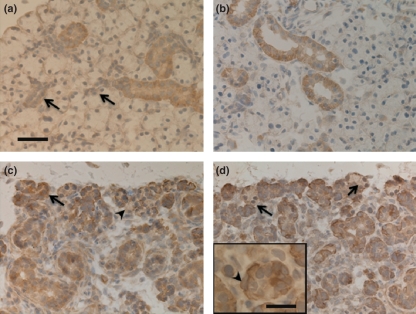
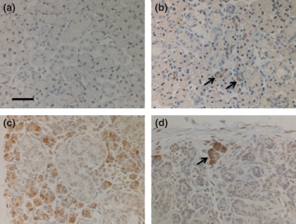
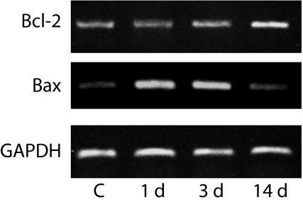
In submandibular gland atrophy, most acinar cells disappear by apoptosis, while many duct cells remain. The present study aimed to establish whether Bcl-2 and Bax, members of the Bcl-2 gene family, regulating the signalling pathway of apoptosis were involved in duct cell survival and acinar cell death in atrophic submandibular glands. The excretory duct of rat submandibular gland was doubly ligated with metal clips from 1 to 14 days to induce atrophy to the gland. The expressions of Bcl-2 and Bax in the atrophic submandibular gland were examined using immunohistochemistry and reverse transcriptase-polymerase chain reaction (RT-PCR). Immunohistochemically, Bcl-2 expression was identified in duct cells in the experimental glands at all time points. Some acinar cells showed Bax positivity 1 day after excretory duct ligation, and there were more Bax-positive acinar cells on days 3 and 5 when many apoptotic acinar cells were observed. Analysis by RT-PCR showed that the expression of mRNA for Bcl-2 became stronger as the glandular atrophy progressed and that Bax mRNA strongly expressed on days 1 and 3. These observations suggest that Bcl-2 inhibits duct cell apoptosis and Bax promotes apoptosis of acinar cells during atrophy of submandibular glands.
Figures
Similar articles
-
Participation of the Fas and Fas ligand systems in apoptosis during atrophy of the rat submandibular glands.Int J Exp Pathol. 2007 Feb;88(1):9-17. doi: 10.1111/j.1365-2613.2006.00511.x. Int J Exp Pathol. 2007. PMID: 17244334 Free PMC article.
-
Epiregulin is critical for the acinar cell regeneration of the submandibular gland in a mouse duct ligation model.J Oral Pathol Med. 2014 May;43(5):378-87. doi: 10.1111/jop.12145. Epub 2013 Dec 20. J Oral Pathol Med. 2014. PMID: 24354788
-
Apoptosis and proliferation of myoepithelial cells in atrophic rat submandibular glands.J Histochem Cytochem. 2001 Dec;49(12):1557-64. doi: 10.1177/002215540104901209. J Histochem Cytochem. 2001. PMID: 11724903
-
Responses of salivary glands to intake of soft diet.J Oral Biosci. 2022 Jun;64(2):210-216. doi: 10.1016/j.job.2022.03.006. Epub 2022 Apr 4. J Oral Biosci. 2022. PMID: 35381373 Review.
-
Apoptotic cell death during regressive changes in salivary glands: A morphological perspective.J Oral Biosci. 2025 Mar;67(1):100585. doi: 10.1016/j.job.2024.100585. Epub 2024 Nov 1. J Oral Biosci. 2025. PMID: 39489340 Review.
Cited by
-
Short-Chain Fatty Acids Alleviate Hepatocyte Apoptosis Induced by Gut-Derived Protein-Bound Uremic Toxins.Front Nutr. 2021 Oct 12;8:756730. doi: 10.3389/fnut.2021.756730. eCollection 2021. Front Nutr. 2021. PMID: 34712690 Free PMC article.
-
Postnatal changes in the development of rat submandibular glands in offspring of diabetic mothers: Biochemical, histological and ultrastructural study.PLoS One. 2018 Oct 10;13(10):e0205372. doi: 10.1371/journal.pone.0205372. eCollection 2018. PLoS One. 2018. PMID: 30304036 Free PMC article.
-
Duct ligation/de-ligation model: exploring mechanisms for salivary gland injury and regeneration.Front Cell Dev Biol. 2024 Jun 25;12:1399934. doi: 10.3389/fcell.2024.1399934. eCollection 2024. Front Cell Dev Biol. 2024. PMID: 38983787 Free PMC article. Review.
-
Identification of drug targets and potential molecular mechanisms for Wantong Jingu Tablet extract in treatment of rheumatoid arthritis: bioinformatics analysis of fibroblast-like synoviocytes.Chin Med. 2020 Jun 5;15:59. doi: 10.1186/s13020-020-00339-5. eCollection 2020. Chin Med. 2020. PMID: 32518584 Free PMC article.
-
[Changes of myoepithelial cells during regeneration of parotid gland].Hua Xi Kou Qiang Yi Xue Za Zhi. 2014 Oct;32(5):446-9. doi: 10.7518/hxkq.2014.05.005. Hua Xi Kou Qiang Yi Xue Za Zhi. 2014. PMID: 25490819 Free PMC article. Chinese.
References
-
- Adams JM, Cory S. The Bcl-2 protein family: arbiters of cell survival. Science. 1998;281:1322–1326. - PubMed
-
- Barnhart BC, Alappat EC, Peter ME. The CD95 type I/type II model. Semin. Immunol. 2003;15:185–193. - PubMed
-
- Green DR. Apoptotic pathways: paper wraps stone blunts scissors. Cell. 2000;102:1–4. - PubMed
-
- Hand AR. Salivary glands. In: Provenza DV, Seibel W, editors. Oral Histology. 2. PA: Lea & Febiger; 1986. pp. 388–417.
-
- Harrison JD, Garrett JR. Mucocele formation in cats by glandular duct ligation. Arch. Oral Biol. 1972;17:1403–1414. - PubMed
MeSH terms
Substances
LinkOut - more resources
Full Text Sources
Research Materials
