Comparative Study
. 2008 Oct;89(5):367-81.
doi: 10.1111/j.1365-2613.2008.00604.x.

Isoproterenol induces primary loss of dystrophin in rat hearts: correlation with myocardial injury

Affiliations
Comparative Study

Isoproterenol induces primary loss of dystrophin in rat hearts: correlation with myocardial injury

Erica C Campos et al. Int J Exp Pathol. 2008 Oct.

Abstract

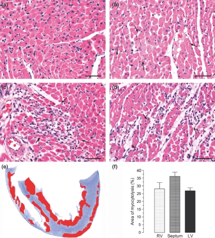
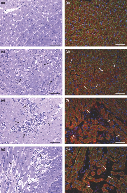
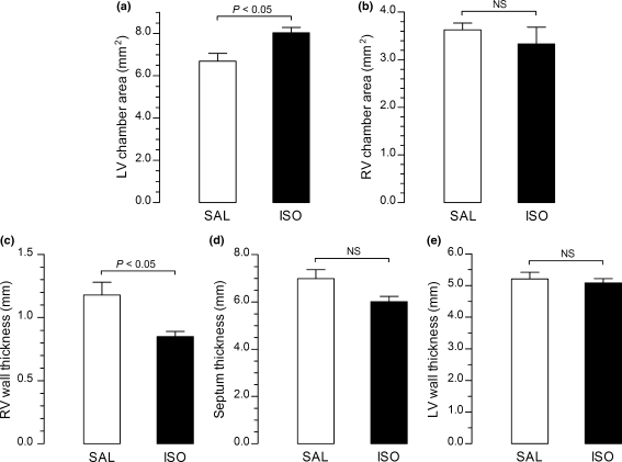
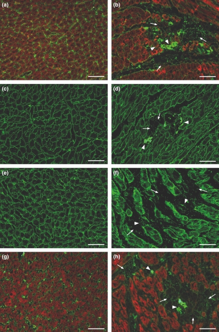
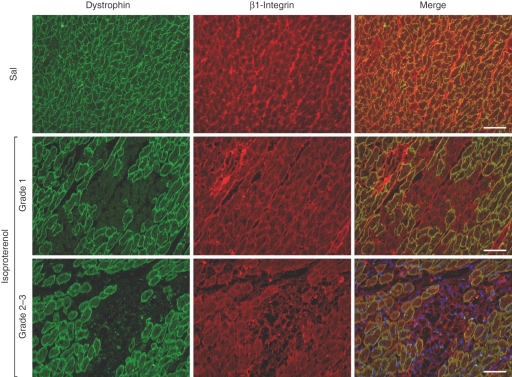
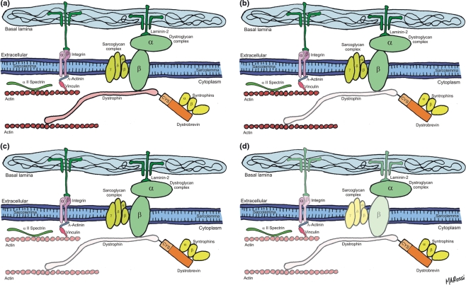
The mechanism of isoproterenol-induced myocardial damage is unknown, but a mismatch of oxygen supply vs. demand following coronary hypotension and myocardial hyperactivity is the best explanation for the complex morphological alterations observed. Severe alterations in the structural integrity of the sarcolemma of cardiomyocytes have been demonstrated to be caused by isoproterenol. Taking into account that the sarcolemmal integrity is stabilized by the dystrophin-glycoprotein complex (DGC) that connects actin and laminin in contractile machinery and extracellular matrix and by integrins, this study tests the hypothesis that isoproterenol affects sarcolemmal stability through changes in the DGC and integrins. We found different sensitivity of the DGC and integrin to isoproterenol subcutaneous administration. Immunofluorescent staining revealed that dystrophin is the most sensitive among the structures connecting the actin in the cardiomyocyte cytoskeleton and the extracellular matrix. The sarcomeric actin dissolution occurred after the reduction or loss of dystrophin. Subsequently, after lysis of myofilaments, gamma-sarcoglycan, beta-dystroglycan, beta1-integrin, and laminin alpha-2 expressions were reduced followed by their breakdown, as epiphenomena of the myocytolytic process. In conclusion, administration of isoproterenol to rats results in primary loss of dystrophin, the most sensitive among the structural proteins that form the DGC that connects the extracellular matrix and the cytoskeleton in cardiomyocyte. These changes, related to ischaemic injury, explain the severe alterations in the structural integrity of the sarcolemma of cardiomyocytes and hence severe and irreversible injury induced by isoproterenol.


Figures

Figure 1
Figure 1
Representative histological features of control myocardium (a) compared with those of myocardium from rats given isoproterenol (85 mg/kg) arranged in an apparent sequence of events : (b) in the first stage, foci of myocytes with tumefaction (arrows), (c) in the second stage, focus of myocytolysis with an inflammatory infiltrated composed of mononuclear cells, and (d) in the third stage, focus showing remnants of cardiomyocytes, empty spaces and a few mononuclear cells. Haematoxylin and eosin. Bar = 50 μm. (e) Schematic drawing showing the mean distribution of necrotic areas in predominantly in the cardiac apex, subendocardium and subepicardium of the left ventricle, in the septum and in the subendocardium of the right ventricle. (f) Mean distribution of the myocytolytic areas in the myocardium of isoproterenol-treated rats.
Figure 3
Figure 3
Representative histological features of control myocardium (a) in comparison with the myocardium of rats given isoproterenol arranged in a sequence of events: (c) foci of myocytes with tumefaction (arrows); (e) focus of myocytolytic necrosis with an inflammatory infiltrate composed of mononuclear cells (arrows); (g) focus showing remnants of myocytes, empty spaces and a few mononuclear cells (arrows). High resolution light microscopy, toluidine blue. Immunofluorescence analysis of dystrophin expression (green fluorescence) in control (b) and isoproterenol-treated myocardium (d, f, h). (d) The immunofluorescent signal is markedly and focally reduced in myocytes of rats given isoproterenol (arrows). (f, h) The foci of myocytolytic necrosis show complete loss of dystrophin immunofluorescent signal with increased number of inflammatory cells (revealed by the blue fluorescence of DAPI) in f (arrows) and a few inflammatory cells in h (arrows). The red fluorescent signal corresponds to actin as stained by phalloidin complexed to Alexa Fluor. Bar = 50 μm.
Figure 2
Figure 2
Graphs showing the mean left and right ventricular chamber areas and right, left and septum thicknesses of hearts from isoproterenol-treated rats (ISO) compared with control values (SAL). The mean area of the left ventricular chamber of the hearts from isoproterenol-treated rats is 20% increased compared with that of controls (a). No significant differences can be seen in the mean area of the right ventricular chamber of isoproterenol-treated rats compared with that of controls (b). The mean right ventricular wall thickness is 26% lower in the isoproterenol-treated rats in comparison with that in controls (c). The mean septum (d) and left ventricular wall thicknesses (e) are similar in both isoproterenol-treated and control groups, although these values tended to be lower in experimental animals.
Figure 4
Figure 4
Representative images of immunofluorescence analysis of γ-sarcoglycan, β-dystroglycan, β-1 integrin and merosin laminin α-2 (green fluorescence) in control (a, c, e, g) and isoproterenol-treated (b, d, f, h) rat hearts. The foci of myocytolytic necrosis show attenuation (arrows) and collapse (arrow heads) of these glycoproteins associated with complete loss of actin (red fluorescence of phalloidin complexed with Alexa Fluor) in the same areas (b, h). Bar = 50 μm.
Figure 5
Figure 5
Sequence of dystrophin and β-1 integrin expression reduction in the same field in saline control and isoproterenol-treated hearts. In the foci corresponding to myocardial lesions type 1, loss of dystrophin expression is clearly seen with the persistence of β-1 integrin. In the foci corresponding to myocardial lesion types 2–3, the loss of dystrophin is associated with attenuation or breakdown of β-1 integrin. Bar = 50 μm.
Figure 6
Figure 6
(a, b) Representative images of the immunofluorescence analysis of macrophages in control and isoproterenol-treated rat hearts (green fluorescence of cytoplasm and blue fluorescence of nuclei). The control myocardium (a) shows no macrophages. The myocardium from isoproterenol-treated rats (b) shows a focal inflammatory infiltrate in the myocytolytic foci characterized by CD-68+ cells. C-E. Representative images of apoptosis assessment. In the control myocardium, the TUNEL reaction was constantly negative (c). In the myocytolytic foci grades 1–3, a positive TUNEL reaction was constantly and clearly noted (arrows) in cardiomyocytes and macrophages (mononuclear cell infiltrate) (d, e). Bar = 50 μm.
Figure 7
Figure 7
(a–c) Representative images of the fluorescence analysis of dystrophin (red fluorescence) and albumin (green fluorescence) in control (a) and isoproterenol-treated rat hearts (b, c). The control myocardium shows uniform dystrophin staining and the albumin restricted to the interstitium just marking the small vessels. The myocardium of rats given isoproterenol shows areas of red and green fluorescence (b) and areas of no dystrophin labelling showing green fluorescence irregularly revealing the cardiomyocytes (c). (d–f) Representative images of fluorescent signals of eNOS (green fluorescence) and actin (red fluorescence) in control (d) and in rats given isoproterenol (e, f). In the myocardium of control animals, the eNOS staining marks the small vessels endothelium. The myocardium from isoproterenol-treated rats shows increased eNOS expression in the small vessels (small arrows in e and arrowheads in f), markedly around the myocytolytic areas and also in the adjacent cytoplasm of myocytes. eNOS expression was practically absent in the myocytolytic areas except for remnants of interstitial small vessels (f, arrows). Bar = 50 μm.
Figure 8
Figure 8
Schematic diagrams representing the sequence of events observed: (a) The DGC and integrins. Dystrophin links actin to the transmembrane proteins dystroglycan and sarcoglycan. Dystroglycan binds to laminin in the extracellular matrix. Integrins also link actin to laminin; (b–d) sequence of events observed: first, loss of dystrophin and presence of sarcomeric actin (b), second, loss of dystrophin and variable degree of sarcomeric actin dissolution (c), and third, loss of dystrophin, sarcomeric actin, and reduction in the expression of γ-sarcoglycan, β-dystroglycan, β1-integrin and laminin α-2 (d).

Similar articles

Cited by

References

    1. Allikian MJ, McNally EM. Processing and assembly of the dystrophin glycoprotein complex. Traffic. 2007;8:177–183. - PubMed
    1. Armstrong SC, Latham CA, Shivell CL, Ganote CE. Ischemic loss of sarcolemmal dystrophin and spectrin: correlation with myocardial injury. J. Mol. Cell. Cardiol. 2001;33:1165–1179. - PubMed
    1. Berges A, van Nassauw L, Timmermans JP, Vrints C. Time-dependent expression pattern of nitric oxide and superoxide after myocardial infarction in rats. Pharmacol. Res. 2007;55:72–79. - PubMed
    1. Berk BC, Fujiwara K, Lehoux S. ECM remodeling in hypertensive heart disease. J. Clin. Invest. 2007;117:568–575. - PMC - PubMed
    1. Chappel CI, Rona G, Balazs T, Gaudry R. Severe myocardial necrosis produced by isoproterenol in the rat. Arch. Int. Pharmacodyn. Ther. 1959;122:123–128. - PubMed

Publication types

MeSH terms