. 2008 Sep 22:5:87.
doi: 10.1186/1742-4690-5-87.

Direct Vpr-Vpr interaction in cells monitored by two photon fluorescence correlation spectroscopy and fluorescence lifetime imaging

Affiliations

Direct Vpr-Vpr interaction in cells monitored by two photon fluorescence correlation spectroscopy and fluorescence lifetime imaging

Joëlle V Fritz et al. Retrovirology. .

Abstract

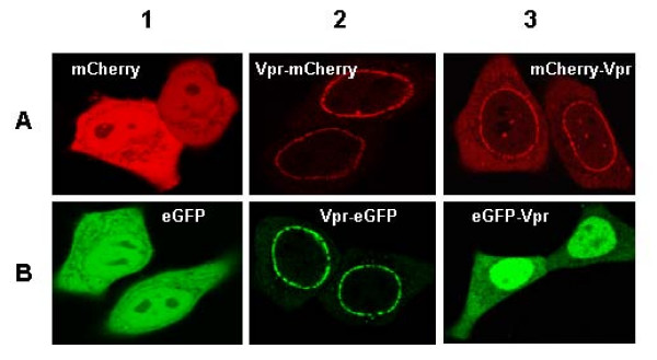
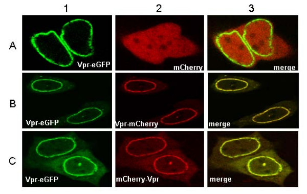
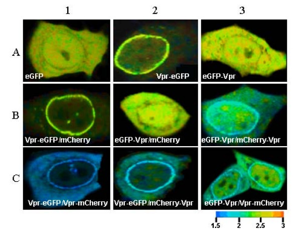
Background: The human immunodeficiency virus type 1 (HIV-1) encodes several regulatory proteins, notably Vpr which influences the survival of the infected cells by causing a G2/M arrest and apoptosis. Such an important role of Vpr in HIV-1 disease progression has fuelled a large number of studies, from its 3D structure to the characterization of specific cellular partners. However, no direct imaging and quantification of Vpr-Vpr interaction in living cells has yet been reported. To address this issue, eGFP- and mCherry proteins were tagged by Vpr, expressed in HeLa cells and their interaction was studied by two photon fluorescence lifetime imaging microscopy and fluorescence correlation spectroscopy.

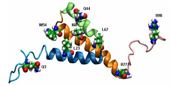
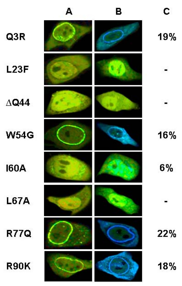
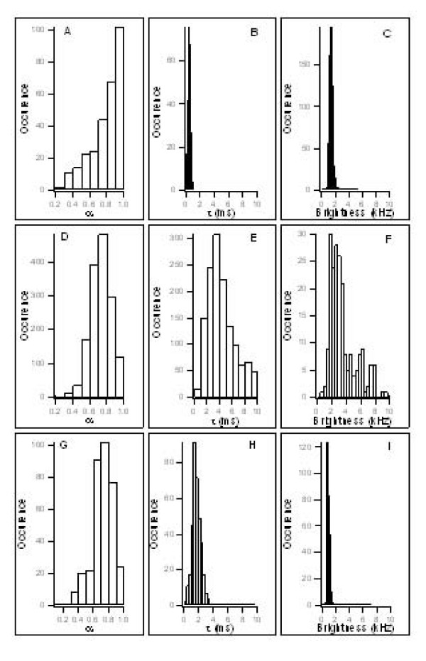
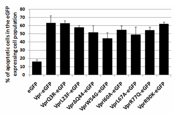
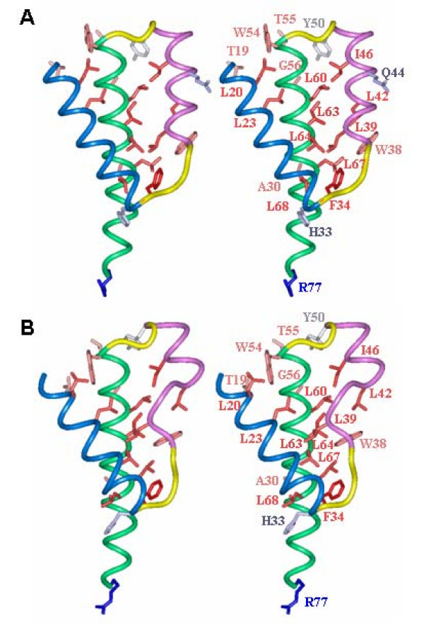
Results: Results show that Vpr forms homo-oligomers at or close to the nuclear envelope. Moreover, Vpr dimers and trimers were found in the cytoplasm and in the nucleus. Point mutations in the three alpha helices of Vpr drastically impaired Vpr oligomerization and localization at the nuclear envelope while point mutations outside the helical regions had no effect. Theoretical structures of Vpr mutants reveal that mutations within the alpha-helices could perturb the leucine zipper like motifs. The DeltaQ44 mutation has the most drastic effect since it likely disrupts the second helix. Finally, all Vpr point mutants caused cell apoptosis suggesting that Vpr-mediated apoptosis functions independently from Vpr oligomerization.

Conclusion: We report that Vpr oligomerization in HeLa cells relies on the hydrophobic core formed by the three alpha helices. This oligomerization is required for Vpr localization at the nuclear envelope but not for Vpr-mediated apoptosis.


Figures

Figure 1
Figure 1
NMR based structure of Vpr. The NMR-based 3D- structure of Vpr (1–96) is characterised by three α helices in close vicinity surrounded by flexible N and C termini [22]. Helices are presented in dark blue (17–33), green (38–50) and orange (54–77). Mutated amino acids Q3R, L23A, ΔQ44, W54G, I60A, L67A, R77Q and R90K are represented in CPK mode. Noticeably, the NMR studies were carried out on the Vpr sequence of the HIV-1 pNL43 strain with a Leucine at the position 60 instead of an Isoleucine for the HIV-1LAI strain used here. Nevertheless, a predictive study on I60 Vpr showed that the third α helix was not altered compared to L60 Vpr (data not shown).
Figure 2
Figure 2
Subcellular localization of eGFP or mCherry tagged Vpr by confocal microscopy. HeLa cells were co-transfected with 0.5 μg of each plasmid and 0.5 μg pcDNA3. Cells were observed by confocal microscopy 24 h post transfection. Each panel shows the major phenotype. (A) mCherry images with excitation at 568 nm and emission at 580 to 700 nm. (B) eGFP images with excitation at 488 nm and emission at 500 to 550 nm. Note the intracellular redistribution of eGFP and mCherry upon fusion with Vpr.
Figure 3
Figure 3
Visualization of the intracellular co-expression eGFP or mCherry tagged Vpr. Plasmid DNA (0.5 μg each) expressing the Vpr fusion proteins were cotransfected in HeLa cells. One day post transfection, images were recorded with an excitation at 488 nm and emission at 500–550 nm to monitor eGFP expression, and with an excitation at 568 nm and emission at 580–700 nm to monitor mCherry expression, respectively. In the merge images, co-localization of the two proteins is indicated in yellow. Each image is representative of the major phenotype. Note the accumulation of the Vpr fusion proteins at or close to the nuclear envelope.
Figure 4
Figure 4
Direct Vpr-Vpr interaction in HeLa cells visualized by FLIM. Cells were transfected with the DNA construct encoding eGFP or eGFP-Vpr alone or in combination with mCherry-Vpr. In the FLIM images, the lifetimes are represented using an arbitrary color scale ranging from blue to red for short and long lifetimes in nanoseconds (right bottom), respectively. The Vpr-eGFP or eGFP-Vpr with short lifetime fluorescence symbolized by the blue color were mainly localized at the nuclear envelope and also in other cell compartments when co transfected with mCherry tagged Vpr. Panels A1 to A3 show the lifetime images of cells expressing eGFP or eGFP-tagged Vpr alone. Panels B1 and B2 represent cells coexpressing eGFP-tagged Vpr and mCherry; Panels B3 and C1-C3 show the lifetime images of cells coexpressing eGFP-tagged Vpr and mCherry-tagged Vpr. Note the accumulation of Vpr fusion proteins at or near the nuclear envelope.
Figure 5
Figure 5
Mapping of Vpr-Vpr interaction by FLIM. HeLa cells were co transfected with mutated Vpr-eGFP and its own counterpart fused to mCherry. FLIM was carried out 24 h posttransfection (see methods). Column A corresponds to the FLIM images of the Vpr-eGFP mutants alone, column B to the FLIM images of cells co expressing the mutant Vpr-eGFP and the mutant Vpr-mCherry. FRET efficiency (E) expressed in percentage represents the average value calculated over the entire cell (column C). The color scale used to create theses images is the same than the one used for figure 4. Note the drastic reduction of Vpr-Vpr interaction and the loss of Vpr nuclear envelope accumulation upon mutating residues L23, Q44, I60 and L67 (column B and C).
Figure 6
Figure 6
Distribution histograms of anomalous diffusion coefficients, diffusion times and count rates/species of eGFP, Vpr-eGFP and ΔQ44 Vpr-eGFP. The anomalous diffusion coefficient (coefficient that accounts for the obstacles encountered by the diffusing species), diffusion times (average time needed to cross the focal volume) and brightness (count rates/species) determined by FCS are expressed as a function of the number of occurrences. A-C correspond to eGFP; D-F correspond to Vpr-eGFP; G-I correspond to ΔQ44 Vpr-eGFP.
Figure 7
Figure 7
Pro-apoptotic properties of the Vpr-eGFP mutants. Cells expressing either the wild type Vpr-eGFP or mutant Vpr-eGFP were selected by fluorescence cytometry, using the eGFP fluorescence. The percentage of cells undergoing apoptosis was assessed by the number of cells labeled with cells with Cy5 alone, or with both Cy5 and PI. Statistical analysis was achieved using the multi-factorial ANOVA test and the Dunnett analysis. Three independent measurements were performed for each assay.
Figure 8
Figure 8
Comparison of the wild type and ΔQ44 Vpr mutant structures. Stereoview of the three dimensional structure of the wild type Vpr determined by NMR (A) and theoretical model for the Vpr ΔQ44 mutant (B). Helices (17–33), (38–50) and (54–77) are represented as ribbon and colored in blue, pink and green, respectively and loops (34–37) and (51–53) are colored in yellow. For clarity, the two disordered extremities of the molecule have not been represented. Residues showing long range correlations on NOESY NMR experiments have been displayed in the stick representation and colored according to their hydrophobicity. Only their side chain atoms have been represented. The network of hydrophobic residues can be observed at the interface of the three α-helices. Note the impact of the ΔQ44 deletion (B) on the partial unfolding of the second helix and the rearrangement of the hydrophobic residues at the interface.

References

    1. Kondo E, Mammano F, Cohen EA, Gottlinger HG. The p6gag domain of human immunodeficiency virus type 1 is sufficient for the incorporation of Vpr into heterologous viral particles. J Virol. 1995;69:2759–2764. - PMC - PubMed
    1. Lavallee C, Yao XJ, Ladha A, Gottlinger H, Haseltine WA, Cohen EA. Requirement of the Pr55gag precursor for incorporation of the Vpr product into human immunodeficiency virus type 1 viral particles. J Virol. 1994;68:1926–1934. - PMC - PubMed
    1. Paxton W, Connor RI, Landau NR. Incorporation of Vpr into human immunodeficiency virus type 1 virions: requirement for the p6 region of gag and mutational analysis. J Virol. 1993;67:7229–7237. - PMC - PubMed
    1. Bachand F, Yao XJ, Hrimech M, Rougeau N, Cohen EA. Incorporation of Vpr into human immunodeficiency virus type 1 requires a direct interaction with the p6 domain of the p55 gag precursor. J Biol Chem. 1999;274:9083–9091. - PubMed
    1. Le Rouzic E, Benichou S. The Vpr protein from HIV-1: distinct roles along the viral life cycle. Retrovirology. 2005;2:11. - PMC - PubMed

Publication types

MeSH terms

Substances

LinkOut - more resources