

The transcription factor ATF3 acts as an oncogene in mouse mammary tumorigenesis
- PMID: 18808719
- PMCID: PMC2564979
- DOI: 10.1186/1471-2407-8-268
The transcription factor ATF3 acts as an oncogene in mouse mammary tumorigenesis
Abstract
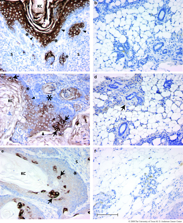
Background: Overexpression of the bZip transcription factor, ATF3, in basal epithelial cells of transgenic mice under the control of the bovine cytokeratin-5 (CK5) promoter has previously been shown to induce epidermal hyperplasia, hair follicle anomalies and neoplastic lesions of the oral mucosa including squamous cell carcinomas. CK5 is known to be expressed in myoepithelial cells of the mammary gland, suggesting the possibility that transgenic BK5.ATF3 mice may exhibit mammary gland phenotypes.
Methods: Mammary glands from nulliparous mice in our BK5.ATF3 colony, both non-transgenic and transgenic, were examined for anomalies by histopathology and immunohistochemistry. Nulliparous and biparous female mice were observed for possible mammary tumor development, and suspicious masses were analyzed by histopathology and immunohistochemistry. Human breast tumor samples, as well as normal breast tissue, were similarly analyzed for ATF3 expression.
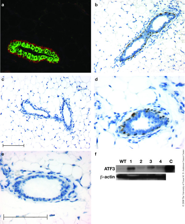
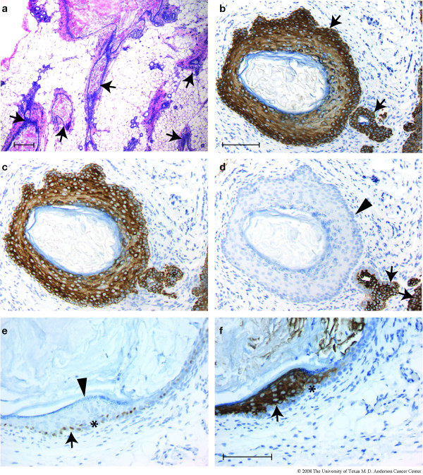
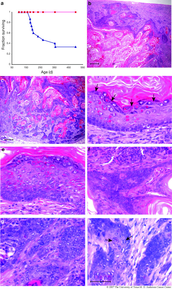
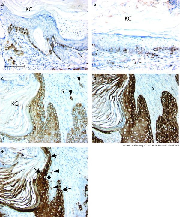
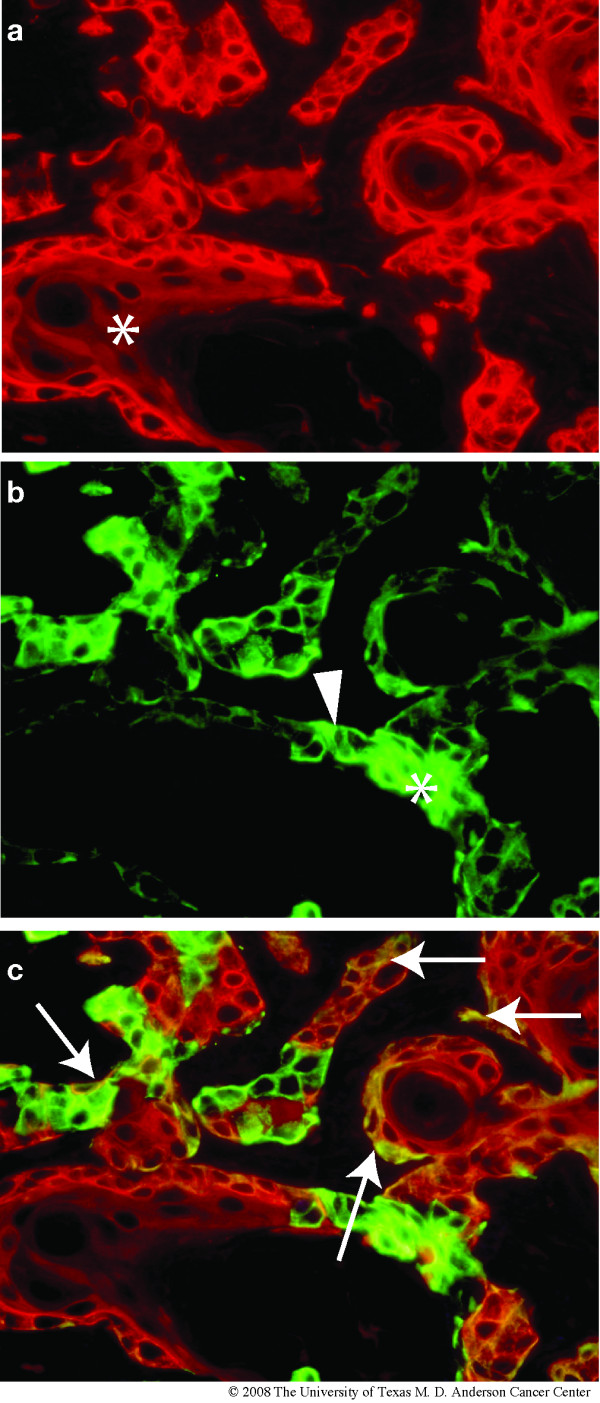
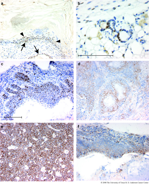
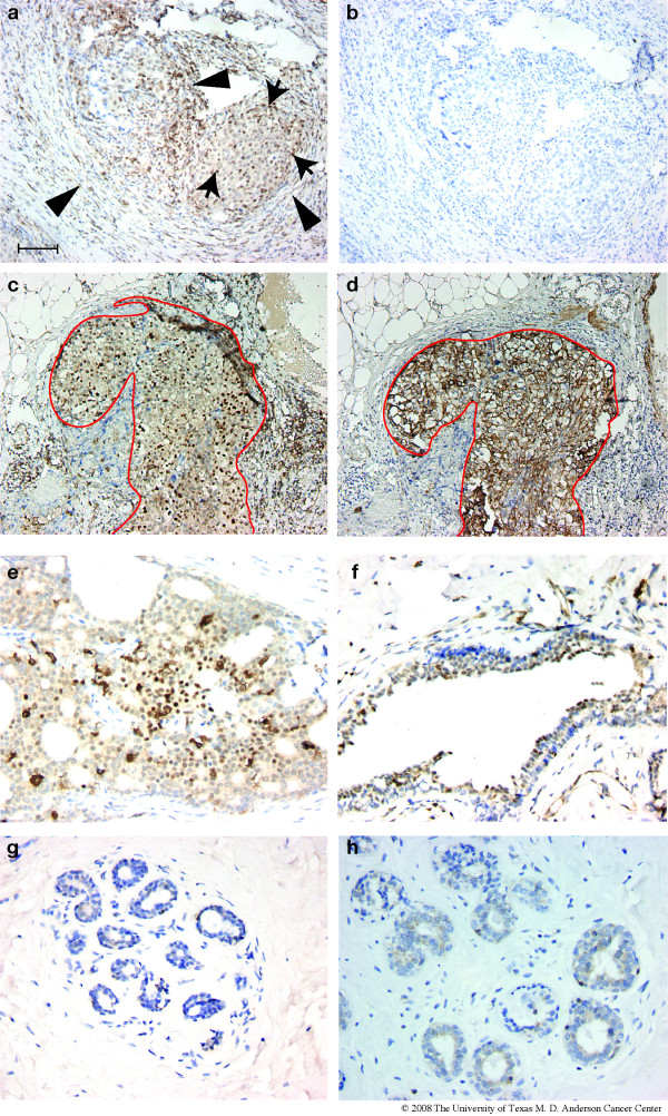
Results: Transgenic BK5.ATF3 mice expressed nuclear ATF3 in the basal layer of the mammary ductal epithelium, and often developed squamous metaplastic lesions in one or more mammary glands by 25 weeks of age. No progression to malignancy was seen in nulliparous BK5.ATF3 or non-transgenic mice held for 16 months. However, biparous BK5.ATF3 mice developed mammary carcinomas with squamous metaplasia between 6 months and one year of age, reaching an incidence of 67%. Cytokeratin expression in the tumors was profoundly disturbed, including expression of CK5 and CK8 (characteristic of basal and luminal cells, respectively) throughout the epithelial component of the tumors, CK6 (potentially a stem cell marker), CK10 (a marker of interfollicular epidermal differentiation), and mIRSa2 and mIRSa3.1 (markers of the inner root sheath of hair follicles). Immunohistochemical studies indicated that a subset of human breast tumors exhibit high levels of nuclear ATF3 expression.
Conclusion: Overexpression of ATF3 in CK5-expressing cells of the murine mammary gland results in the development of squamous metaplastic lesions in nulliparous females, and in mammary tumors in biparous mice, suggesting that ATF3 acts as a mammary oncogene. A subset of human breast tumors expresses high levels of ATF3, suggesting that ATF3 may play an oncogenic role in human breast tumorigenesis, and therefore may be useful as either a biomarker or therapeutic target.
Figures
Similar articles
-
Epidermal hyperplasia and oral carcinoma in mice overexpressing the transcription factor ATF3 in basal epithelial cells.Mol Carcinog. 2007 Jun;46(6):476-87. doi: 10.1002/mc.20298. Mol Carcinog. 2007. PMID: 17295236
-
Local over-expression of prolactin in differentiating mouse mammary gland induces functional defects and benign lesions, but no carcinoma.J Endocrinol. 2006 Aug;190(2):271-85. doi: 10.1677/joe.1.06829. J Endocrinol. 2006. PMID: 16899561
-
ATF3-Induced Mammary Tumors Exhibit Molecular Features of Human Basal-Like Breast Cancer.Int J Mol Sci. 2021 Feb 26;22(5):2353. doi: 10.3390/ijms22052353. Int J Mol Sci. 2021. PMID: 33652981 Free PMC article.
-
Transgenic mice reveal roles for TGFalpha and EGF receptor in mammary gland development and neoplasia.J Mammary Gland Biol Neoplasia. 1997 Apr;2(2):119-29. doi: 10.1023/a:1026347629876. J Mammary Gland Biol Neoplasia. 1997. PMID: 10882298 Review.
-
The biology of mammary transgenes: five rules.J Mammary Gland Biol Neoplasia. 1996 Jan;1(1):61-73. doi: 10.1007/BF02096303. J Mammary Gland Biol Neoplasia. 1996. PMID: 10887481 Review.
Cited by
-
The antioxidant transcription factor Nrf2 modulates the stress response and phenotype of malignant as well as premalignant pancreatic ductal epithelial cells by inducing expression of the ATF3 splicing variant ΔZip2.Oncogene. 2019 Feb;38(9):1461-1476. doi: 10.1038/s41388-018-0518-3. Epub 2018 Oct 9. Oncogene. 2019. PMID: 30302023
-
DNA methylation level of promoter region of activating transcription factor 5 in glioma.J Zhejiang Univ Sci B. 2015 Sep;16(9):757-62. doi: 10.1631/jzus.B1500067. J Zhejiang Univ Sci B. 2015. PMID: 26365117 Free PMC article.
-
ATF3 transcription factor and its emerging roles in immunity and cancer.J Mol Med (Berl). 2009 Nov;87(11):1053-60. doi: 10.1007/s00109-009-0520-x. Epub 2009 Aug 25. J Mol Med (Berl). 2009. PMID: 19705082 Free PMC article. Review.
-
Up-Regulation of Activating Transcription Factor 3 in Human Fibroblasts Inhibits Melanoma Cell Growth and Migration Through a Paracrine Pathway.Front Oncol. 2020 Apr 21;10:624. doi: 10.3389/fonc.2020.00624. eCollection 2020. Front Oncol. 2020. PMID: 32373541 Free PMC article.
-
Loss of activating transcription factor 3 prevents KRAS-mediated pancreatic cancer.Oncogene. 2021 Apr;40(17):3118-3135. doi: 10.1038/s41388-021-01771-z. Epub 2021 Apr 16. Oncogene. 2021. PMID: 33864001 Free PMC article.
References
-
- Perou CM, Sorlie T, Eisen MB, Rijn M van de, Jeffrey SS, Rees CA, Pollack JR, Ross DT, Johnsen H, Akslen LA, Fluge O, Pergamenschikov A, Williams C, Zhu SX, Lonning PE, Borresen-Dale AL, Brown PO, Botstein D. Molecular portraits of human breast tumours. Nature. 2000;406:747–752. doi: 10.1038/35021093. - DOI - PubMed
Publication types
MeSH terms
Substances
Grants and funding
LinkOut - more resources
Full Text Sources
Molecular Biology Databases
Research Materials
Miscellaneous
