

Review
doi: 10.1016/j.cbpa.2008.08.008.
Super-resolution microscopy by nanoscale localization of photo-switchable fluorescent probes
Affiliations
- PMID: 18809508
- PMCID: PMC2642911
- DOI: 10.1016/j.cbpa.2008.08.008
Item in Clipboard
Review
Super-resolution microscopy by nanoscale localization of photo-switchable fluorescent probes
Curr Opin Chem Biol.
2008 Oct.
Abstract
A new form of super-resolution fluorescence microscopy has emerged in recent years, based on the high accuracy localization of individual photo-switchable fluorescent labels. Image resolution as high as 20 nm in the lateral dimensions and 50 nm in the axial direction has been attained with this concept, representing an order of magnitude improvement over the diffraction limit. The demonstration of multicolor imaging with molecular specificity, three-dimensional (3D) imaging of cellular structures, and time-resolved imaging of living cells further illustrates the exciting potential of this method for biological imaging at the nanoscopic scale.
Figures
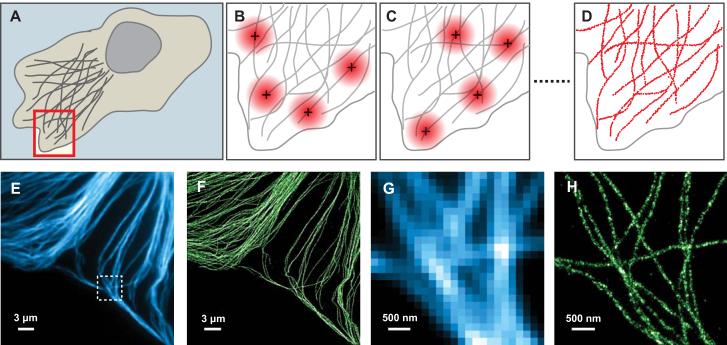
Super-resolution imaging by high-precision localization of photo-switchable fluorophores. (A - D) The imaging concept. (A) Schematic of a cell in which the structure of interest (grey filaments in this case) are labeled with photo-switchable fluorophores (not shown). All fluorophores are initially in the non-fluorescent state. The red box indicates the area shown in panels B - D. (B) An activation cycle: a sparse set of fluorophores are activated to the fluorescent state, such that their images (large red circles) do not overlap. The image of each fluorophore appears as a diffraction-broadened spot, and the position of each activated fluorophore is determined by fitting to find the centroid of the spot (black crosses). (C) A subsequent activation cycle: a different set of fluorophores are activated and their positions are determined as before. (D) After a sufficient number of fluorophores have been localized, a high resolution image is constructed by plotting the measured positions of the fluorophores (red dots). The resolution of this image is not limited by diffraction, but by the accuracy of each fluorophore localization and by the number of fluorophore positions obtained. (E - H) Comparison of conventional immunofluorescence images of microtubules in a BSC1 cell (E, G) and STORM images (F, H) of the same areas. In the STORM images, each localization is rendered as a Gaussian peak whose width corresponds to the theoretical localization accuracy. The areas shown in (G) and (H) are expanded views of the region defined by the dashed box in (E). The microtubules are immuno-labeled with photo-switchable Cy3 - Alexa 647 dye pairs. Panels E - H reproduced from [50] with permission.
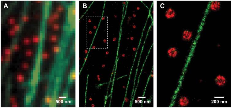
Multi-color STORM imaging. (A, B) A comparison of two-color conventional immunofluoresence (A) and STORM (B) images of microtubules (green) and clathrin-coated pits (red) in fixed BSC1 cells. The antibodies used for microtubule staining were labeled with Cy2 and Alexa 647 (a structural analog of Cy5) as the activator and reporter, respectively. For clathrin labeling the antibodies were labeled with Cy3 and Alexa 647. (C) A further magnified view of the boxed region shown in (B). Panels B and C reproduced from [50] with permission.
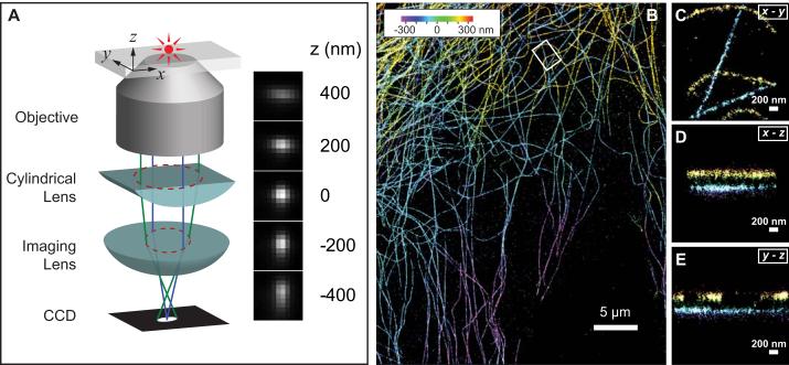
Three-dimensional STORM imaging. (A) Simplified optical diagram illustrating the principle of determining the z coordinate of a fluorescent object from the ellipticity of its image by introducing a cylindrical lens into the imaging path. The right panel shows images of a fluorophore at various z positions. (B) Three-dimensional STORM image of microtubules in a cell. The z-position information is color-coded according to the color scale bar. (C - E) The x-y, x-z, and y-z cross sections of a small region of the cell outlined by the white box in (B), showing five microtubule filaments. Figure reproduced from [53] with permission.
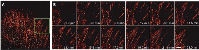
Live-cell PALM imaging (A) PALM image of a wide area of the cell, showing elongated and punctate adhesion complexes in a CHO cell expressing paxillin fused to EosFP. Scale bar 5 μm. (B) Higher magnification of the green boxed region in (A) illustrating adhesion complex initiation and elongation over a time course of 23 minutes. Scale bar 3 μm. Figure reproduced from [61] with permission.
References
-
- Giepmans BN, Adams SR, Ellisman MH, Tsien RY. The fluorescent toolbox for assessing protein location and function. Science. 2006;312:217–224. - PubMed
-
- Koster AJ, Klumperman J. Electron microscopy in cell biology: integrating structure and function. Nat Rev Mol Cell Biol. 2003;(Suppl):SS6–10. - PubMed
-
- Abbe E. Beitrage zur Theorie des Mikroskops und der mikroskopischen Wahrnehmung. Arch Mikroskop Anat. 1873;9:413–420.
-
- Pawley JB, editor. Handbook of Biological Confocal Microscopy. Springer; New York: 2006.
-
- Helmchen F, Denk W. Deep tissue two-photon microscopy. Nat Methods. 2005;2:932–940. - PubMed
Publication types
MeSH terms
Substances
Grants and funding
LinkOut - more resources
Full Text Sources
Other Literature Sources
