. 2008 Dec;28(23):7245-58.
doi: 10.1128/MCB.01085-08. Epub 2008 Sep 22.

Genotoxic stress-induced cyclin D1 phosphorylation and proteolysis are required for genomic stability

Affiliations

Genotoxic stress-induced cyclin D1 phosphorylation and proteolysis are required for genomic stability

Laura L Pontano et al. Mol Cell Biol. 2008 Dec.

Abstract

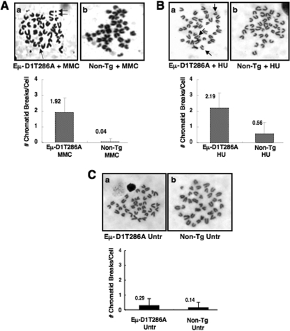
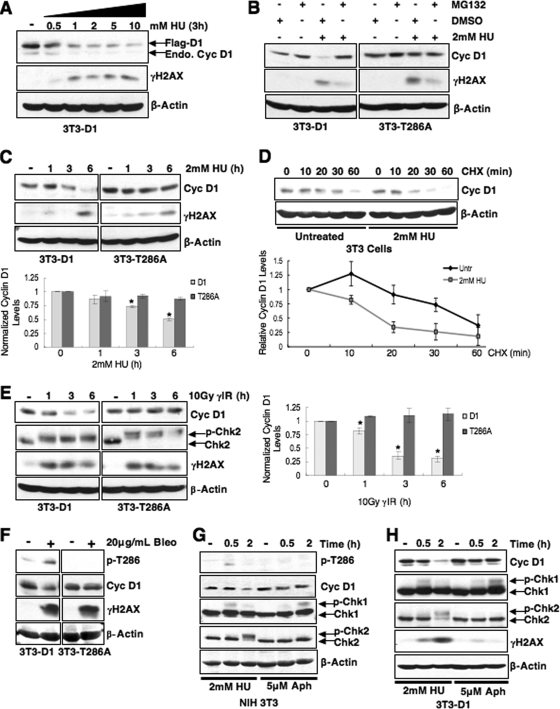
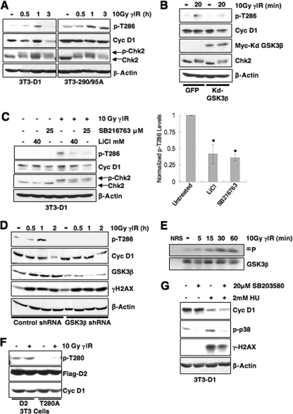
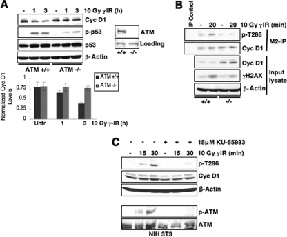
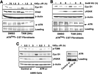
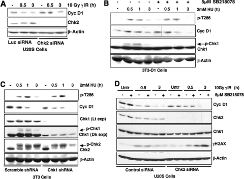
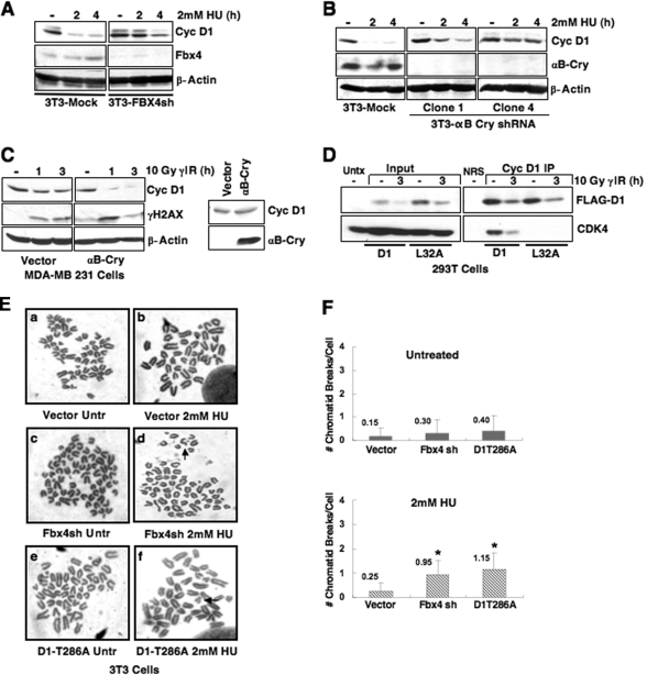
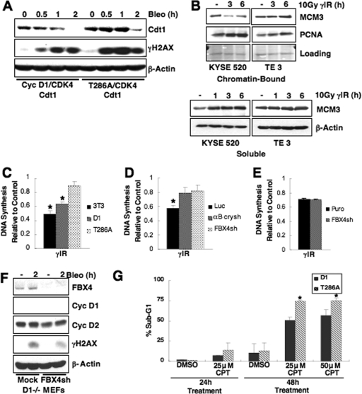
While mitogenic induction of cyclin D1 contributes to cell cycle progression, ubiquitin-mediated proteolysis buffers this accumulation and prevents aberrant proliferation. Because the failure to degrade cyclin D1 during S-phase triggers DNA rereplication, we have investigated cellular regulation of cyclin D1 following genotoxic stress. These data reveal that expression of cyclin D1 alleles refractory to phosphorylation- and ubiquitin-mediated degradation increase the frequency of chromatid breaks following DNA damage. Double-strand break-dependent cyclin D1 degradation requires ATM and GSK3beta, which in turn mediate cyclin D1 phosphorylation. Phosphorylated cyclin D1 is targeted for proteasomal degradation after ubiquitylation by SCF(Fbx4-alphaBcrystallin). Loss of Fbx4-dependent degradation triggers radio-resistant DNA synthesis, thereby sensitizing cells to S-phase-specific chemotherapeutic intervention. These data suggest that failure to degrade cyclin D1 compromises the intra-S-phase checkpoint and suggest that cyclin D1 degradation is a vital cellular response necessary to prevent genomic instability following genotoxic insult.


Figures

FIG. 1.
FIG. 1.
(A and B) Chromatid breaks following MMC (A) or HU (B) treatment occur at a greater frequency in splenocytes expressing Eμ-D1T286A. Splenocytes derived from nontransgenic or premalignant Eμ-D1T286A mice were cultured for 24 h in the presence of 100 μM MMC or cultured for 24 h, followed by a 2-h pulse with 2 mM HU and resuspension in fresh media. Cells were then treated with Colcemid for 2 h to allow arrest in metaphase. (C) Untreated nontransgenic and Eμ-D1T286A splenocytes do not exhibit a significant number of chromatid breaks. Representative images of metaphase spreads are shown, and the average number of chromatid breaks per metaphase spread was quantified for each treatment.
FIG. 2.
FIG. 2.
Phosphorylation-dependent proteolysis of cyclin D1 following DSB induction. (A) Cyclin D1 is degraded following replication stress. Synchronous 3T3-D1 cells were treated with increasing doses of HU as indicated, and cell lysates were probed for cyclin D1 and γH2AX as a marker of DSB induction. The 2 mM dose was selected for further HU experiments. (B and C) Cyclin D1 degradation is phosphorylation and proteasome dependent. (B) Synchronous 3T3-D1- or 3T3-D1T286A-expressing cells were treated with MG132 and HU for 4 h; immunoblots were performed as in panel A. (C) 3T3-D1 or D1T286A cells were treated with HU as indicated, and cyclin D1 levels were assessed. (D) HU treatment decreases cyclin D1 t1/2. S-phase NIH 3T3 cells were pretreated with HU, followed by 100 μg of cycloheximide (CHX)/ml as indicated, and cyclin D1 protein levels were assessed. (E) Cyclin D1 levels rapidly decline following γIR. 3T3-D1 or 3T3-D1T286A cells were subjected to 10Gy γIR, and cyclin D1 levels were assessed. Chk2 mobility shift (upper band) and γH2AX served as markers of DSBs. (F) The DSB-inducing agent bleomycin promotes cyclin D1 degradation. Asynchronous 3T3-D1 or D1T286A cells were treated with 20 μg of bleomycin (Bleo)/ml for 2 h, and the cyclin D1 levels were assessed. (G and H) Stalled DNA replication is not sufficient to induce cyclin D1 proteolysis. Synchronous NIH 3T3 cells (G) or NIH 3T3 cells stably expressing ectopic cyclin D1 (H) were treated with 2 mM HU to induce DSBs or 5 μM aphidicolin (Aph) to stall DNA replication without inducing DNA breaks. Immunoblot for Chk1 mobility shift served as a marker of stalled replication, and a Chk2 mobility shift indicates DSB induction. Cyclin D1 is phosphorylated (G) and degraded (G and H) following DSB induction.
FIG. 3.
FIG. 3.
IR-induced degradation is specific to wild-type cyclin D1 protein. (A and B) Cyclin D1 is refractory to degradation in esophageal carcinoma cells harboring the endogenous P287A mutation. Asynchronously proliferating cells expressing wild-type cyclin D1 (KYSE 520) or P287A mutant cyclin D1 (TE3/TE7) were irradiated with a moderate dose (A) or high dose (B), and cyclin D1 levels and checkpoint activation were assessed by immunoblotting as indicated. (C) Cyclin D1, but not cyclin D2 or cyclin E, is degraded following DNA damage. Asynchronously proliferating NIH 3T3 cells stably expressing cyclin D1 were irradiated, followed by recovery at 37°C. Protein stability was assessed by direct Western blotting.
FIG. 4.
FIG. 4.
GSK3β catalyzes rapid cyclin D1 T286 phosphorylation following DNA damage. (A) Cyclin D1 phosphorylation is induced by DNA damage. Asynchronous 3T3-D1, 3T3-D1-290/95A, or 3T3-D1T286A were subjected to γIR, and immunoblots were performed with antibodies specific to p-T286, cyclin D1, and γH2AX. (B) Expression of kdGSK3β abrogates cyclin D1 phosphorylation following γIR. Lysates NIH 3T3 cells transiently expressing green fluorescent protein (GFP) or kdGSK3β plus GFP and γ-irradiated were blotted for p-T286, cyclin D1, and myc to detect myc-tagged kdGSK3β. (C) Inhibition of GSK3β attenuates cyclin D1 phosphorylation following DNA damage. 3T3-D1 cells were pretreated with the GSK3β inhibitor LiCl or SB216763, followed by γIR and recovery for 30 min. Cyclin D1 phosphorylation was assessed by immunoblot for p-T286 and total cyclin D1. Chk2 mobility shift and γH2AX indicate DSB induction. (D) shRNA-mediated knockdown of GSK3β attenuates cyclin D1 phosphorylation. 293T cells were transfected with control or GSK3β-specific shRNA, cyclin D1, and CDK4 constructs. Cells were subjected to γIR and recovery for the times indicated, and cyclin D1 phosphorylation, efficiency of GSK3β knockdown, and checkpoint activation were assessed by immunoblotting. (E) GSK3β is activated following DNA damage. Endogenous GSK3β precipitated from cells following γIR was utilized as a kinase for in vitro reactions with recombinant myelin basic protein substrate. (F) Cyclin D2 Thr-280 phosphorylation is not induced following DNA damage. NIH 3T3 cells were transiently transfected with wild-type or D2T280A. Cells were irradiated 48 h posttransfection; cyclin D2 phosphorylation was assessed by Western blotting, and probing for total flag-tagged cyclin D2 served as a loading and transfection control. (G) Cyclin D1 is rapidly degraded following DNA damage in the presence of p38 inhibitor. Synchronous NIH 3T3 cells were pretreated with 20 μM SB203580 or DMSO for 30 min, followed by HU treatment for 2.5 h. Cell lysates were prepared and probed for p-p38 and cyclin D1.
FIG. 5.
FIG. 5.
ATM signaling is required for cyclin D1 phosphorylation following DSB induction. (A) Cyclin D1 degradation is attenuated in cells lacking ATM expression. Immortalized ATM+/+ or ATM−/− mouse embryonic fibroblasts (MEFs) expressing Flag-tagged cyclin D1 were gamma irradiated, and cyclin D1 levels were assessed. p-p53/total p53 induction served as markers of checkpoint activation in these cell lines. (B) DNA damage-induced cyclin D1 phosphorylation is only detected in cells expressing ATM. Immortalized ATM+/+ or ATM−/− MEFs expressing Flag-tagged cyclin D1 were irradiated as indicated, and lysates were precipitated with M2-agarose to detect Flag-tagged protein. Cyclin D1 phosphorylation and total levels were assessed by immunoblotting. (C) ATM signaling is required for cyclin D1 phosphorylation. NIH 3T3 cells were pretreated with DMSO or the specific ATM inhibitor KU-55933 for 30 min, followed by γIR and recovery for 15 or 30 min. Phosphorylated and total cyclin D1 were detected by immunoblotting. A second Western blot was performed with these lysates to probe for phosphorylated ATM and total ATM to ensure efficacy of ATM inhibition.
FIG. 6.
FIG. 6.
ATR is dispensable for DSB-induced cyclin D1 loss. (A and B) ATR deletion does not alter cyclin D1 degradation following DNA damage. ATRflox/− CET fibroblasts were treated with 4-OH tamoxifen (TAM) for 24 h, followed by incubation at 37°C for 12 h to allow sufficient ATR depletion. Cells were then irradiated (A) or treated with 2 mM HU (B) as indicated, and the cyclin D1 levels were assessed by immunoblotting. Significant ATR deletion was obtained by TAM treatment. (C) siRNA-mediated attenuation of ATR expression does not inhibit cyclin D1 phosphorylation or degradation. U20S cells were transfected with ATR-specific siRNA and irradiated as indicated 60 h after siRNA delivery. Cyclin D1 phosphorylation and total cyclin D1 and the efficiency of ATR knockdown were assessed by immunoblotting.
FIG. 7.
FIG. 7.
Examining the role of Chk1 and Chk2 kinases in cyclin D1 regulation. (A) Chk2 knockdown modestly attenuates DNA damage-induced cyclin D1 degradation. U20S cells were transiently transfected with Chk2-specific or luciferase control siRNAs. At 60 h posttransfection, the cells were gamma-irradiated, and cyclin D1 levels were assessed. (B and C) Chk1 activation is not required for cyclin D1 phosphorylation. Synchronous 3T3-D1 cells were pretreated with the Chk1 inhibitor SB218078 for 30 min (B), or NIH 3T3 cells were transduced with Chk1 shRNA lentivirus (C). Cells were treated with HU, and cyclin D1 phosphorylation or total protein levels were assessed by immunoblotting. (D) Chk1 and Chk2 activity is not synergistic in regulating cyclin D1. U20S cells expressing Chk2-specific siRNA were treated with the Chk1 inhibitor SB218078. Cyclin D1 levels were assessed by immunoblotting following DNA damage.
FIG. 8.
FIG. 8.
The SCFFbx4-αBcrystallin E3 ubiquitin ligase regulates cyclin D1 stability following DNA damage. Fbx4 and αB crystallin are required for efficient cyclin D1 degradation following DNA damage. (A and B) Synchronous NIH 3T3 cells stably expressing control and either Fbx4 (A) or αB crystallin-specific shRNAs (B) were treated with 2 mM HU, and cell lysates were probed for Fbx4 (A) or αB crystallin (B) to confirm knockdown and cyclin D1 to assess protein stability following DNA damage. (C) Asynchronous MDA-MB231 cells expressing empty vector or αB crystallin were γ-irradiated, followed by recovery at 37°C, and cyclin D1 stability was assessed by immunoblotting. Confirmation of exogenous αB crystallin expression in this cell line is shown. (D) Mutation of leucine 32 within the RxxL motif does not alter DNA damage-induced cyclin D1 degradation. 293T cells (chosen for high transfection efficiency and expression of SCF-Fbx4 ligase components) were transfected with Flag-tagged wild-type or D1L32A, along with CDK4 and GFP. Cell lysates were prepared and precipitated with M2 agarose beads to recognize Flag-tagged protein. Immunoprecipitates and input Western blots were probed with cyclin D1 and CDK4 antibodies to assess cyclin D1 and CDK4 association. (E) Disruption of SCFFbx4-αBcrystallin sensitizes cells to genomic instability after HU treatment. NIH 3T3 cells stably expressing vector control or Fbx4-specific shRNA were treated with 2 mM HU for 3 h, followed by nocodazole treatment to arrest cells in metaphase. Cells were fixed, and metaphase spreads were prepared. Representative images of chromatid breaks are shown (E), and the average number of chromatid breaks per spread was quantified for each treatment (F). NIH 3T3 cells stably expressing cyclin D1T286A served as a positive control for the presence of chromatid breaks following HU.
FIG. 9.
FIG. 9.
Failure to degrade cyclin D1 compromises the intra-S-phase checkpoint response to DNA damage and sensitizes cells to CPT. (A) The replication factor Cdt1 is stabilized following DNA damage in cells expressing cyclin D1T286A. HeLa cells were transfected with wild-type or cyclin D1T286A, CDK4, and Cdt1. Cells were treated with 10 μg of bleomycin/ml as indicated, and Cdt1 levels were assessed by immunoblotting. (B) Cyclin D1 stabilization promotes maintenance of MCM proteins on chromatin. Esophageal carcinoma cell lines expressing endogenous wild-type or D1P287A were irradiated and harvested at the time points indicated. Cell lysates were fractionated into chromatin-bound and soluble fractions; MCM3 binding to chromatin was assessed by immunoblotting on chromatin-bound fractions. Ponceau-S stain served as a control for equal loading of chromatin-bound fractions, and PCNA is a representative chromatin-bound protein. (C) Expression of D1T286A results in RDS. RDS assays were performed in synchronous parental NIH 3T3 cells or 3T3-D1 or 3T3-D1T286A cells. Equal concentrations of DNA were counted for [3H] and [14C]thymidine incorporation. DNA synthesis, calculated as the ratio of [3H] to [14C], was normalized to the nonirradiated control for each cell line. (D) Disruption of SCFFbx4-αBcrystallin promotes RDS. RDS assays were performed as in panel C utilizing cells stably expressing luciferase, αB crystallin, or Fbx4-specific shRNA. (E) Fbx4 attenuation in cyclin D1-null cells does not result in RDS. RDS assays as in panel C were performed in wild-type or cyclin D1-null MEFs stably expressing empty vector or Fbx4-specific shRNAs. (F) Fbx4 levels are attenuated in cyclin D1-null MEFs stably expressing Fbx4-specific shRNA. (G) Failure to degrade cyclin D1 sensitizes cells to the CPT. 3T3-D1 or D1T286A cells were treated with CPT as indicated for 24 or 48 h. Cells were fixed and stained with propidium iodide and subjected to FACS analysis; a sub-G1 population is indicative of apoptotic cells.

References

    1. Agami, R., and R. Bernards. 2000. Distinct initiation and maintenance mechanisms cooperate to induce G1 cell cycle arrest in response to DNA damage. Cell 10255-66. - PubMed
    1. Aggarwal, P., M. D. Lessie, D. I. Lin, L. Pontano, A. B. Gladden, B. Nuskey, A. Goradia, M. A. Wasik, A. J. Klein-Szanto, A. K. Rustgi, C. H. Bassing, and J. A. Diehl. 2007. Nuclear accumulation of cyclin D1 during S phase inhibits Cul4-dependent Cdt1 proteolysis and triggers p53-dependent DNA rereplication. Genes Dev. 212908-2922. - PMC - PubMed
    1. Alt, J. R., J. L. Cleveland, M. Hannink, and J. A. Diehl. 2000. Phosphorylation-dependent regulation of cyclin D1 nuclear export and cyclin D1-dependent cellular transformation. Genes Dev. 143102-3114. - PMC - PubMed
    1. Barbash, O., P. Zamfirova, D. I. Lin, X. Chen, K. Yang, H. Nakagawa, F. Lu, A. K. Rustgi, and J. A. Diehl. 2008. Mutations in Fbx4 inhibit dimerization of the SCF(Fbx4) ligase and contribute to cyclin D1 overexpression in human cancer. Cancer Cell 1468-78. - PMC - PubMed
    1. Barnes, D. M., and C. E. Gillett. 1998. Cyclin D1 in breast cancer. Breast Cancer Res. Treat. 521-15. - PubMed

Publication types

LinkOut - more resources