. 2008 Dec;28(23):7001-11.
doi: 10.1128/MCB.00732-08. Epub 2008 Sep 22.

Redundant roles of SMAD2 and SMAD3 in ovarian granulosa cells in vivo

Affiliations

Redundant roles of SMAD2 and SMAD3 in ovarian granulosa cells in vivo

Qinglei Li et al. Mol Cell Biol. 2008 Dec.

Abstract

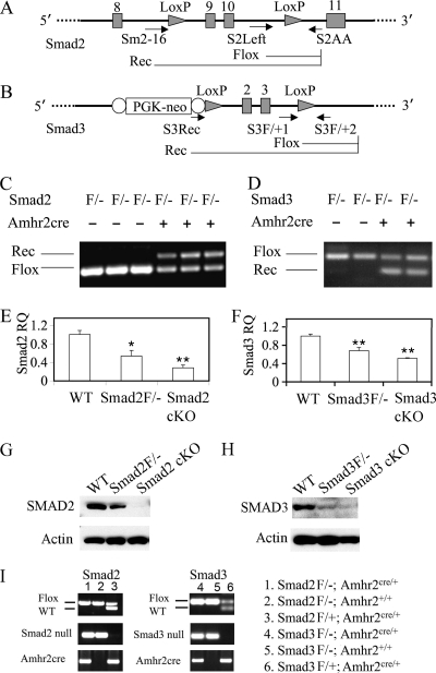
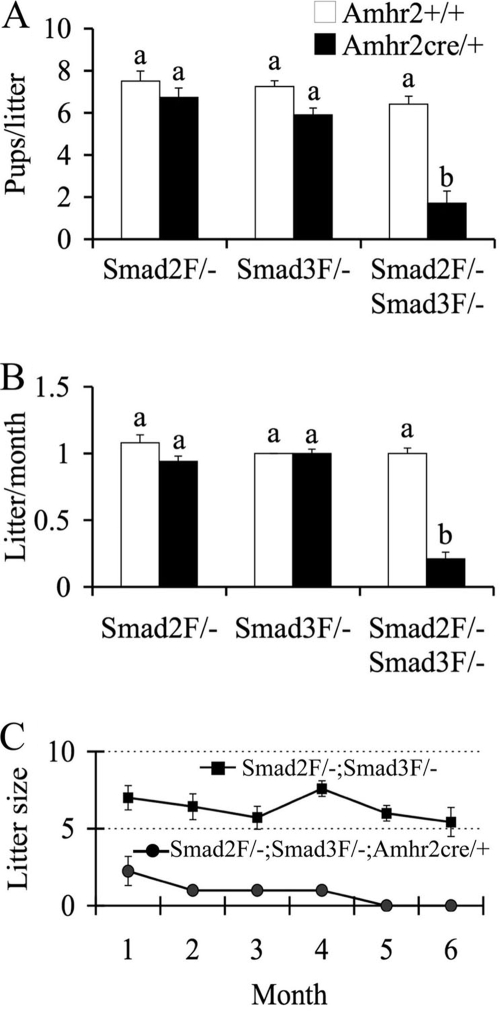
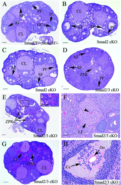
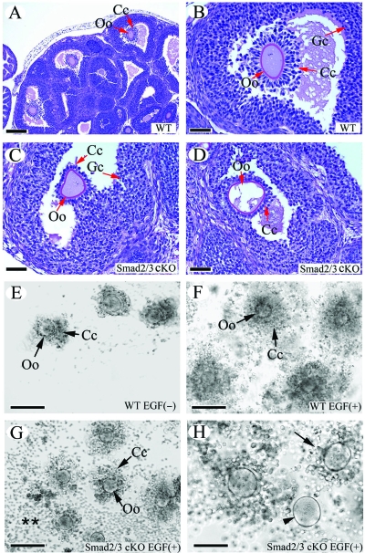
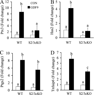
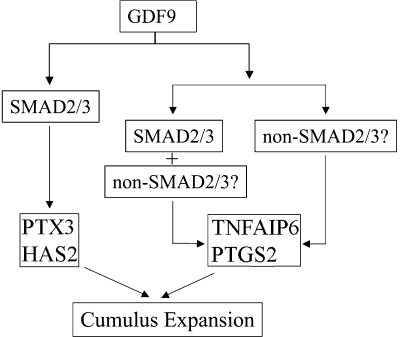
Transforming growth factor beta (TGF-beta) superfamily members are critical in maintaining cell growth and differentiation in the ovary. Although signaling of activins, TGF-betas, growth differentiation factor 9, and nodal converge preferentially to SMAD2 and SMAD3, the in vivo functions and redundancy of these SMADs in the ovary and female reproduction remain largely unidentified. To circumvent the deleterious phenotypic aspects of ubiquitous deletion of Smad2 and Smad3, a conditional knockout strategy was formulated to selectively inactivate Smad2, Smad3, or both Smad2 and Smad3 in ovarian granulosa cells. While granulosa cell ablation of individual Smad2 or Smad3 caused insignificant changes in female fertility, deletion of both Smad2 and Smad3 led to dramatically reduced female fertility and fecundity. These defects were associated with the disruption of multiple ovarian processes, including follicular development, ovulation, and cumulus cell expansion. Furthermore, the impaired expansion of cumulus cells may be partially associated with altered cumulus expansion-related transcripts that are regulated by SMAD2/3 signaling. Our results indicate that SMAD2 and SMAD3 function redundantly in vivo to maintain normal female fertility and further support the involvement of an intraovarian SMAD2/3 pathway in mediating oocyte-produced signals essential for coordinating key events of the ovulatory process.


Figures

FIG. 1.
FIG. 1.
Construction and recombination of Smad2 and Smad3 floxed alleles. (A) Schematic representation of Smad2 conditional allele with exons 9 and 10 (shaded vertical bars) flanked by two loxP sites (shaded triangles). Primers S2Left and S2AA detect the Smad2 floxed allele, while Sm2-16 and S2AA identify the recombined allele of Smad2. Real-time PCR primers were designed based on the nucleotide sequence of exon 10. (B) Schematic representation of Smad3 conditional allele with exons 2 and 3 (shaded vertical bars) flanked by two loxP sites (shaded triangles). A PGK promoter driven neomycin (PGK-neo) cassette flanked by flip recombinase consensus sequences (open ovals) was illustrated. Primers S3F/+1 and S3F/+2 detect the Smad3 floxed allele, while S3Rec and S3F/+2 detect the recombined allele. Quantitative PCR primers were designed based on the nucleotide sequence of exon 1 (forward primer) and exon 2 (reverse primer; deleted exon) of Smad3. (C and D) Recombination of Smad2 and Smad3 floxed alleles in the genomic DNA of granulosa cells. Note that the recombined Smad2 or Smad3 allele was only detectable in mice that express Amhr2cre recombinase. (E and F) Relative Smad2 and Smad3 mRNA level in granulosa cells of control and Smad2 or Smad3 cKO mice. The relative mRNA abundance of genes relative to WT controls was determined by using the ΔΔCT method. The data are shown as means ± the SEM. *, P < 0.05l **, P < 0.01 (versus WT controls). (G and H) Representative Western blots of total SMAD2 and SMAD3 protein levels in the granulosa cells of control and cKO mice. Compared to WT mice, SMAD2 (∼58 kDa) was markedly reduced (data not shown) or became undetectable in the independent pools of granulosa cells from Smad2 cKO mice (G). SMAD3 protein (50 kDa) was reduced to background levels in the Smad3 cKO mice versus WT controls (H). (I) Genotyping of Smad2 or Smad3 cKO mice using genomic PCR. Representative PCR images are shown on the left, and the corresponding genotypes are denoted on the right. Flox/− and Flox/+ are abbreviated as F/− and F/+, respectively.
FIG. 2.
FIG. 2.
Fertility changes in Smad2, Smad3, and Smad2/3 cKO mice during a 6-month testing period. (A) Litter size of Smad2, Smad3, and Smad2/3 cKO mice versus controls. Smad2 cKO (n = 6) and Smad3 cKO (n = 8) did not show significant differences in the litter size versus Smad2flox/−; Amhr2+/+ (n = 6) and Smad3flox/−; Amhr2+/+ (n = 7) controls. The litter size of the Smad2/3 cKO mice (n = 7) was dramatically reduced compared to Smad2flox/−; Smad3flox/−; Amhr2+/+ controls (n = 8). (B) Litters/month in Smad2, Smad3, and Smad2/3 cKO mice versus controls. The litter/month of Smad2flox/−; Amhr2cre/+ and Smad3flox/−; Amhr2cre/+ mice did not significantly differ from the controls. However, the litters/month were dramatically decreased in Smad2/3 cKO mice compared to the controls. (C) Time course of litter sizes in Smad2/3 cKO mice during a 6-month testing period. The Smad2/3 cKO mice became infertile after 4 months of breeding. The data are shown as means ± the SEM, and bars without a common superscript are significantly different at P < 0.01. Flox/− is abbreviated as F/−.
FIG. 3.
FIG. 3.
Histological analyses of ovaries from Smad2/3 cKO mice. (A) Ovarian histology of a 3-month-old Smad2flox/−; Smad3flox/− mouse. The ovary contained follicles at all developmental stages (primordial, primary, secondary, and antral follicles) and corpora lutea. Flox/− is abbreviated as F/−. (B) Ovarian histology of a 3-month-old Smad2 cKO mouse demonstrating normal follicular development. (C) Ovarian histology of an 8-month-old Smad2 cKO mouse demonstrating indiscernible abnormalities. (D, E, and F) Ovarian histology of 3-month-old Smad2/3 cKO mice. Note that the ovaries of Smad2/3 cKO mice contained fewer antral follicles (D and E) compared to the controls (A). Other histological features of Smad2/3 cKO ovaries were the accumulation of ZPRs (D to F) and the presence of luteinizing follicles (F; arrow) with trapped oocytes (F; arrowhead). (G) Ovarian histology of an 8-month-old Smad2/3 cKO mouse. Note the lack of large antral follicles, the accumulation of ZPRs, and the presence of a luteinizing follicle. (H) Ovarian histology of an 8-month-old Smad2/3 cKO mouse demonstrating aberrant cumulus histology (arrow). PF, primordial and primary follicle; SF, secondary follicle; AF, antral follicle; CL, corpora lutea; GC, granulosa cell; Cc, cumulus cell; TC, theca cell; Oo, oocyte; LF, luteinizing follicle. Scale bars: A to E and G, 200 μm; F, 100 μm; H, 50 μm.
FIG. 4.
FIG. 4.
In vivo and in vitro cumulus expansion defects in Smad2/3 cKO mice. (A and B) Cumulus expansion in WT mice treated with PMSG-hCG. Note the outward radiation pattern of the expanded cumulus cells from the oocytes illustrated in panel B. (C and D) The cumulus cells failed to undergo expansion in response to PMSG-hCG treatment in Smad2/3 cKO mice. Note that fewer cumulus cells were attached to the oocyte, and these cells did not undergo the typical expansion observed in control mice (A and B). Even though expansion of cumulus cells was observed occasionally in some large antral follicles, the expansion pattern observed in WT mice was lacking (data not shown). (E) COCs from WT mice cultured in the absence of EGF. (F) In vitro expansion of cumulus cells from WT mice stimulated by EGF (10 ng/ml). Note the expansion pattern of cumulus cells outward from the oocytes in the COC culture. (G and H) Cultured COCs from Smad2/3 cKO mice stimulated by EGF (10 ng/ml). Note the impaired expansion of cumulus cells (H; arrow) and the denuded oocyte (H; arrowhead) in the COC culture. The detached cumulus cells from COCs are indicated (**). Oo, oocyte; Cc, cumulus cell; Gc, granulosa cell. Scale bars: A, 200 μm; B to D and H, 50 μm; E to G, 100 μm.
FIG. 5.
FIG. 5.
Effect of conditional deletion of Smad2 and Smad3 on GDF9-stimulated cumulus expansion-related transcript expression in granulosa cells. Immature female mice (WT, n = 5; Smad2/3 cKO [S2/3 cKO], n = 3) were primed with PMSG, and granulosa cells were collected and cultured in the absence or presence of 100 ng of recombinant mouse GDF9/ml. GDF9 significantly induced expression of Ptx3 (A), Has2 (B), Ptgs2 (C), and Tnfaip6 (D) (4- to 8-fold increases) in the granulosa cells of WT mice. However, in the granulosa cells of Smad2/3 cKO mice, GDF9-stimulated Ptx3 (A) and Has2 (B) increases were suppressed. GDF9-stimulated Ptgs2 (C) and Tnfaip6 (D) increases still occurred in the Smad2/3 cKO granulosa cells but in an attenuated pattern. Changes in the relative mRNA expression of genes relative to WT control were determined by using the ΔΔCT method. The data are shown as means ± the SEM, and bars without a common superscript are significantly different.
FIG. 6.
FIG. 6.
Hypothetical working model for GDF9 induction of cumulus expansion-related transcripts through SMAD2/3-dependent and/or SMAD2/3-independent pathways. GDF9 can signal through SMAD2/3 to regulate PTX3 and HAS2, since conditional deletion of Smad2 and Smad3 substantially suppressed GDF9 induced Ptx3 and Has2 expression. In contrast, GDF9-induced Ptgs2 and Tnfaip6 expression still occurred in Smad2/3 conditionally deleted cells, suggesting that GDF9 might signal through both SMAD2/3-dependent and/or SMAD2/3-independent pathways to confer the regulation of these genes. The nature of the SMAD-independent pathways remains to be identified.

Similar articles

Cited by

References

    1. Andreu-Vieyra, C., R. Chen, and M. M. Matzuk. 2007. Effects of granulosa cell-specific deletion of Rb in Inha-alpha null female mice. Endocrinology. 1483837-3849. - PubMed
    1. Andreu-Vieyra, C., R. Chen, and M. M. Matzuk. 2008. Conditional deletion of the retinoblastoma (Rb) gene in ovarian granulosa cells leads to premature ovarian failure. Mol. Endocrinol. 222141-2161. - PMC - PubMed
    1. Ashcroft, G. S., S. J. Mills, K. C. Flanders, L. A. Lyakh, M. A. Anzano, S. C. Gilliver, and A. B. Roberts. 2003. Role of Smad3 in the hormonal modulation of in vivo wound healing responses. Wound Repair Regen. 11468-473. - PubMed
    1. Billiar, R. B., J. B. St Clair, N. C. Zachos, M. G. Burch, E. D. Albrecht, and G. J. Pepe. 2004. Localization and developmental expression of the activin signal transduction proteins Smads 2, 3, and 4 in the baboon fetal ovary. Biol. Reprod. 70586-592. - PubMed
    1. Boerboom, D., M. Paquet, M. Hsieh, J. Liu, S. P. Jamin, R. R. Behringer, J. Sirois, M. M. Taketo, and J. S. Richards. 2005. Misregulated Wnt/β-catenin signaling leads to ovarian granulosa cell tumor development. Cancer Res. 659206-9215. - PubMed

Publication types

LinkOut - more resources