. 2008 Dec;28(23):7199-211.
doi: 10.1128/MCB.01040-08. Epub 2008 Sep 22.

MCAK-independent functions of ch-Tog/XMAP215 in microtubule plus-end dynamics

Affiliations

MCAK-independent functions of ch-Tog/XMAP215 in microtubule plus-end dynamics

Alexis R Barr et al. Mol Cell Biol. 2008 Dec.

Abstract

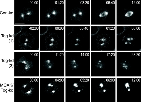
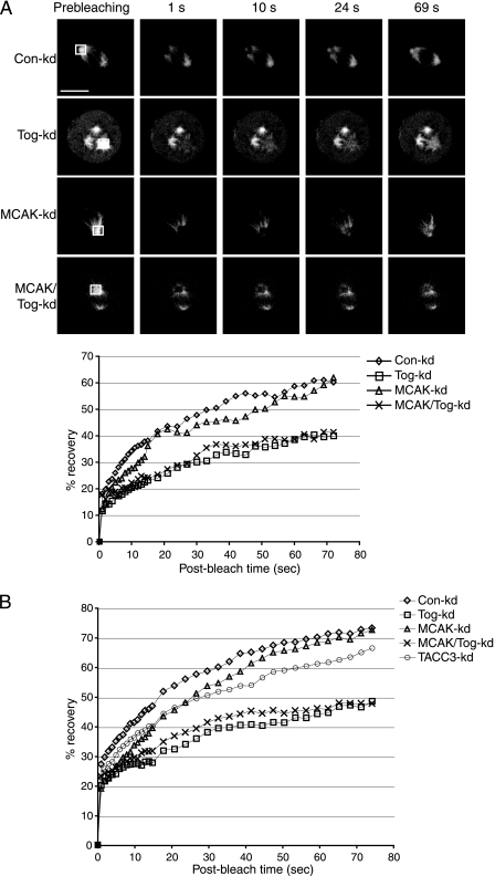
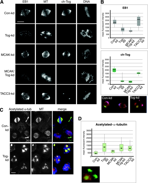
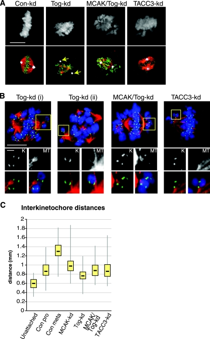
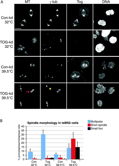
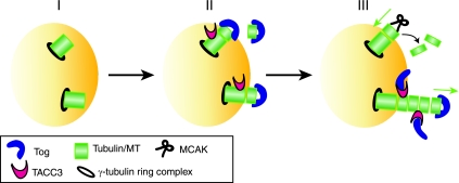
The formation of a functional bipolar mitotic spindle is essential for genetic integrity. In human cells, the microtubule polymerase XMAP215/ch-Tog ensures spindle bipolarity by counteracting the activity of the microtubule-depolymerizing kinesin XKCM1/MCAK. Their antagonistic effects on microtubule polymerization confer dynamic instability on microtubules assembled in cell-free systems. It is, however, unclear if a similar interplay governs microtubule behavior in mammalian cells in vivo. Using real-time analysis of spindle assembly, we found that ch-Tog is required to produce or maintain long centrosomal microtubules after nuclear-envelope breakdown. In the absence of ch-Tog, microtubule assembly at centrosomes was impaired and microtubules were nondynamic. Interkinetochore distances and the lengths of kinetochore fibers were also reduced in these cells. Codepleting MCAK with ch-Tog improved kinetochore fiber length and interkinetochore separation but, surprisingly, did not rescue centrosomal microtubule assembly and microtubule dynamics. Our data therefore suggest that ch-Tog has at least two distinct roles in spindle formation. First, it protects kinetochore microtubules from depolymerization by MCAK. Second, ch-Tog plays an essential role in centrosomal microtubule assembly, a function independent of MCAK activity. Thus, the notion that the antagonistic activities of MCAK and ch-Tog determine overall microtubule stability is too simplistic to apply to human cells.


Figures

FIG. 1.
FIG. 1.
Tog-kd cells develop multipolar spindles after NEBD. Shown are still images from movies following spindle assembly in siRNA-treated synchronized HeLa cells expressing GFP-tubulin. Time 00:00 (minutes:seconds) indicates NEBD. The images in the top row follow a Con-kd cell from NEBD to assembly of the bipolar spindle. Two examples are shown for multipolar-spindle assembly in Tog-kd cells: row Tog-kd (1) depicts aster formation near existing poles (the “splitting” phenotype), while row Tog-kd (2) shows de novo aster formation at random sites. Note that spindle pole splitting occurs almost simultaneously with NEBD in Tog-kd (1). Supernumerary poles move toward and then cluster around the centrosomes in Tog-kd (2). The images in the bottom row show how a small aster appears and moves away from the spindle pole in MCAK/Tog-kd cells before being incorporated into a bipolar spindle. Scale bar = 10 μm.
FIG. 2.
FIG. 2.
ch-Tog is required for efficient recovery of GFP-tubulin signal at spindle poles following photobleaching. (A) Examples of GFP-tubulin recovery after photobleaching of the framed areas (shown in the prebleaching column). The graph shows mean signal intensities in the bleach window plotted against time for individual cells from the images. Tog-kd and MCAK/Tog-kd cells display similar recovery patterns that are clearly distinct from the recovery patterns of Con-kd and MCAK-kd cells. Scale bar = 10 μm. (B) Averaged signal intensities from 10 cells per siRNA treatment. The distribution of the data is summarized in a box plot in Fig. S3 in the supplemental material.
FIG. 3.
FIG. 3.
MTs in Tog-kd spindles are long lived, and their growing tips do not cluster around centrosomes. (A) EB1 signal is reduced in the spindle poles of Tog-kd and MCAK/Tog-kd cells. The cells were immunostained with anti-EB1, Cy3-anti-β-tubulin/MT (MT), and anti-ch-Tog antibodies. DNA was stained with Hoechst stain. Scale bar = 10 μm. (B) Box plots representing distributions of mean fluorescence intensities of anti-EB1 (top) and anti-ch-Tog (bottom) antibody staining at the spindle pole area (see Materials and Methods for details). Examples of spindle pole area selections in Con-kd and Tog-kd cells are highlighted in various colors on projected images in which MTs are stained with Cy3-anti-β-tubulin (red). Note that as these areas encompass the minus-end region of the spindle, TACC3-kd selections contain reduced mean fluorescence intensities of anti-ch-Tog, which is consistent with a decrease in ch-Tog levels on spindle MTs in these cells. The horizontal lines indicate median values. The data represent the spindle poles of 20 cells per siRNA treatment processed within a single experiment. Similar distributions were obtained in three independent experiments. (C) Acetylated-α-tubulin (α-tub) levels are elevated in Tog-kd spindles. In prometaphase, anti-acetylated-α-tubulin antibodies stain Tog-kd spindles more strongly than Con-kd spindles. Anti-acetylated α-tubulin is shown in green, Cy3-anti-β-tubulin/MT in red, and DNA in blue in the merged images. Note that a significant proportion of MTs are acetylated in Tog-kd cells. (D) Box plot representing the distribution of mean fluorescence intensities of anti-acetylated-α-tubulin staining in the spindles of 30 cells. Similar results were obtained in three independent experiments. An example of area selection in a Tog-kd cell is highlighted in red on the projected image, in which MTs are stained with Cy3-anti-β-tubulin (green). Note that examples of selections are shown on projected images, but all calculations were carried out in three-dimensional volumes built of Z stacks. Scale bar = 10 μm.
FIG. 4.
FIG. 4.
Kinetochore MTs are abundant in Tog-kd cells, but they neither associate with the centrosome nor provide tension at kinetochores. (A) Kinetochore MTs were visualized in siRNA-treated cells following treatment with calcium extraction buffer. Centrosomes associate with few or no calcium-stable MTs in Tog-kd cells (arrows). Codepleting MCAK with ch-Tog rescued this phenotype. The panels on the left show DNA staining. In the merged images (bottom row), centrosomes are stained with anti-CDK5RAP2 antibodies (white), kinetochores with anti-Hec1 antibodies (green), and MTs with Cy3-anti-β-tubulin (red). Scale bar = 10 μm. (B) Close-ups of chromosome attachments in siRNA-treated cells. The cells were treated with calcium extraction buffer prior to fixation. Kinetochores are marked with anti-Hec1 (green) and MTs with Cy3-anti-β-tubulin (red) antibodies. DNA staining is in blue in the merged images. The large panels (scale bar = 10 μm) show individual cells, whereas the images (scale bar = 1 μm) arranged below depict magnifications of the areas framed in yellow. The black-and-white images are close-ups of either kinetochores (K) or MTs (MT), while the merged images below show the same areas without (left) or with (right) DNA staining. The images are projections of four confocal Z slices (0.3-μm steps). The two chromosomes shown in Tog-kd (i) have monopolar attachments, but kinetochore MTs are weak in both cases. The chromosome in Tog-kd (ii) shows syntelic-like attachment. Note that the kinetochores display side-on instead of end-on attachments to MTs, a common phenotype in these cells. Monopolar attachments remain frequent in MCAK/Tog-kd cells, but kinetochore MTs are more pronounced there than in Tog-kd cells. Monopolar attachments are also present in TACC3-kd cells. (C) Box plot representation of the distribution of interkinetochore distances in various siRNA-treated cells. Unattached chromosomes were scored in prophase Con-kd cells. Prometaphase (Con pro) and metaphase (Con meta) Con-kd cells were scored separately. In Tog-kd, TACC3-kd, and Tog/MCAK-kd cells, only kinetochores of chromosomes present on metaphase-like plates were included. The data represent 200 kinetochore pairs (10 pairs per cell) for each category.
FIG. 5.
FIG. 5.
Chromatin-dependent MT assembly is important to maintain spindle morphology and MT polymer levels in Tog-kd cells. (A) tsBN2 cells treated with control or ch-Tog siRNAs are shown at permissive (32°C) and restrictive (39.5°C) temperatures. The cells were incubated for 4 h at the restrictive temperature before fixation. At 39.5°C, the spindle morphology radically changes in Tog-kd cells. The cell marked with an asterisk is devoid of ch-Tog and MT polymer. Note that extra poles are visible in the lower Con-kd cell at 39.5°C. MTs were detected using Cy3-anti-β-tubulin antibodies and centrosomes with anti-γ-tubulin antibodies. Scale bar = 10 μm. (B) Graph representing quantitation of mitotic-spindle phenotypes in tsBN2 cells at 32°C and 39.5°C. Specific examples for each category are highlighted in panel A with arrows of matching colors. The data were collected from 100 mitotic cells (n = 3). The error bars correspond to standard deviations.
FIG. 6.
FIG. 6.
Schematic model explaining how TACC3 and ch-Tog function at the plus ends of nascent MTs. (I) Short MT polymers are nucleated off the γ-tubulin ring complex. (II) ch-Tog initiates MT growth by binding tubulin heterodimers (1, 55) and loading them onto the plus ends of short MTs (7). In the absence of ch-Tog, centrosomal MT plus ends do not grow. TACC3 can bind MTs independently of ch-Tog (21). (III) MT-bound TACC3 interacts with ch-Tog. This interaction could perform a dual role by (i) promoting the rate of MT elongation by maintaining ch-Tog in the proximity of plus ends and (ii) inducing a conformational change in ch-Tog that allows effective recruitment of heterodimers into the MT lattice. TACC3 and ch-Tog therefore cooperate to achieve rapid polymerization of plus ends. The plus ends of fast-growing MTs (with ch-Tog/TACC3) may be less amenable to depolymerization by MCAK than slowly growing ones (without ch-Tog/TACC3).

References

    1. Al-Bassam, J., N. A. Larsen, A. A. Hyman, and S. C. Harrison. 2007. Crystal structure of a TOG domain: conserved features of XMAP215/Dis1-family TOG domains and implications for tubulin binding. Structure 15355-362. - PubMed
    1. Andrews, P. D., Y. Ovechkina, N. Morrice, M. Wagenbach, K. Duncan, L. Wordeman, and J. R. Swedlow. 2004. Aurora B regulates MCAK at the mitotic centromere. Dev. Cell 6253-268. - PubMed
    1. Barr, A. R., and F. Gergely. 2007. Aurora-A: the maker and breaker of spindle poles. J. Cell Sci. 1202987-2996. - PubMed
    1. Barros, T. P., K. Kinoshita, A. A. Hyman, and J. W. Raff. 2005. Aurora A activates D-TACC-Msps complexes exclusively at centrosomes to stabilize centrosomal microtubules. J. Cell Biol. 1701039-1046. - PMC - PubMed
    1. Bastiaens, P., M. Caudron, P. Niethammer, and E. Karsenti. 2006. Gradients in the self-organization of the mitotic spindle. Trends Cell Biol. 16125-134. - PubMed

Publication types