. 2008 Dec;28(23):7126-38.
doi: 10.1128/MCB.00897-08. Epub 2008 Sep 22.

PML activates transcription by protecting HIPK2 and p300 from SCFFbx3-mediated degradation

Affiliations

PML activates transcription by protecting HIPK2 and p300 from SCFFbx3-mediated degradation

Yutaka Shima et al. Mol Cell Biol. 2008 Dec.

Abstract

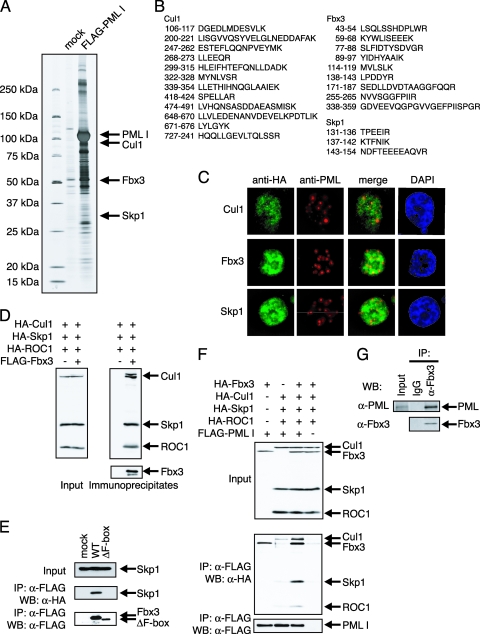
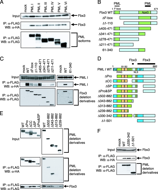
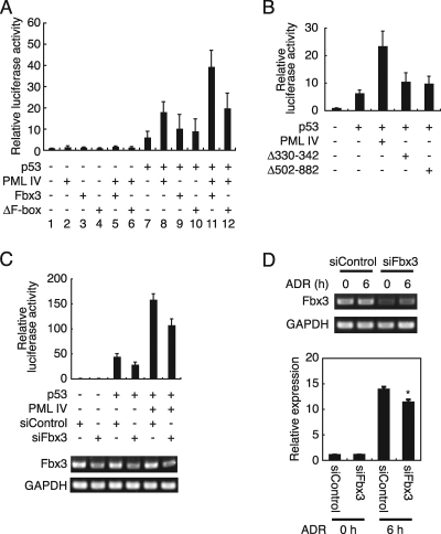
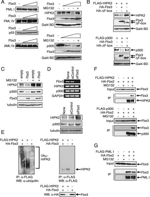
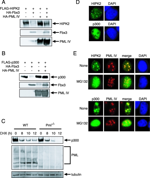
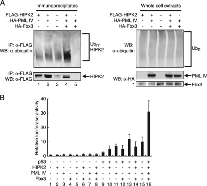
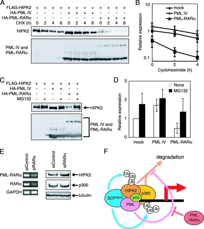
PML, a nuclear protein, interacts with several transcription factors and their coactivators, such as HIPK2 and p300, resulting in the activation of transcription. Although PML is thought to achieve transcription activation by stabilizing the transcription factor complex, little is known about the underlying molecular mechanism. To clarify the role of PML in transcription regulation, we purified the PML complex and identified Fbxo3 (Fbx3), Skp1, and Cullin1 as novel components of this complex. Fbx3 formed SCF(Fbx3) ubiquitin ligase and promoted the degradation of HIPK2 and p300 by the ubiquitin-proteasome pathway. PML inhibited this degradation through a mechanism that unexpectedly did not involve inhibition of the ubiquitination of HIPK2. PML, Fbx3, and HIPK2 synergistically activated p53-induced transcription. Our findings suggest that PML stabilizes the transcription factor complex by protecting HIPK2 and p300 from SCF(Fbx3)-induced degradation until transcription is completed. In contrast, the leukemia-associated fusion PML-RARalpha induced the degradation of HIPK2. We discuss the roles of PML and PML-retinoic acid receptor alpha, as well as those of HIPK2 and p300 ubiquitination, in transcriptional regulation and leukemogenesis.


Figures

FIG. 1.
FIG. 1.
Ubiquitin ligase SCFFbx3 is part of the PML complex. (A) Purification of the PML complex. The PML complex was purified from cell lysates prepared from K562 cells carrying an empty vector (mock) or stably expressing FLAG-tagged PML I. The complexes were incubated with anti-FLAG antibody-conjugated agarose, and the bound materials were eluted with the FLAG peptide. The eluates were resolved by SDS-PAGE and visualized by silver staining. The proteins were identified by mass spectrometry. (B) The amino acid sequences of the peptides derived from the fractions specific to the FLAG-PML I-expressing cells. The proteins in the specific fractions were identified as Cul1, Fbx3, and Skp1. (C) Cul1, Fbx3, and Skp1 colocalized with PML I. MCF7 cells were cotransfected with pLNCX-FLAG-PML I and pcDNA-HA-Cul1, pcDNA-HA-Fbx3, or pcDNA-HA-Skp1. Cul1, Fbx3, and Skp1 were stained with anti-HA antibody and PML I was stained with anti-PML (001) antibody. DAPI, 4′,6′-diamidino-2-phenylindole. (D) Fbx3 forms a complex with Skp1, Cul1, and ROC1. BOSC23 cells were transfected with pcDNA-HA-Skp1, pcDNA-HA-Cul1, pcDNA-HA-ROC1, and either the empty vector (−) or pcDNA-FLAG-Fbx3. The expression of Skp1, Cul1, and ROC1 in the lysates of transfectants was detected by immunoblotting using anti-HA antibody (left). The Fbx3 complex was immunoprecipitated with anti-FLAG antibody. The immunoprecipitates were analyzed by immunoblotting using anti-HA and anti-FLAG antibodies (right). (E) The F-box domain is required for the interaction between Fbx3 and Skp1. BOSC23 cells were transfected with pcDNA-HA-Skp1 and either mock or pcDNA-FLAG-Fbx3 constructs as indicated. The expression of Skp1 in the lysates of transfectants was detected by immunoblotting using anti-HA antibody (top). The lysates of transfectants were incubated with anti-FLAG antibody. The immunoprecipitates were analyzed by immunoblotting using anti-HA (middle) and anti-FLAG (bottom) antibodies. (F) PML interacts with SCFFbx3 through Fbx3. BOSC23 cells were cotransfected with pcDNA-HA-Fbx3, pcDNA-HA-Skp1, pcDNA-HA-Cul1, pcDNA-HA-ROC1, and either mock empty vector or pLNCX-FLAG-PML I. The interactions between PML I and components of SCFFbx3 were analyzed as described for panel D. (G) Endogenous Fbx3 interacts with endogenous PML. SKNO-1 cells were lysed and Fbx3 was immunoprecipitated with anti-Fbx3 antibody. The immunoprecipitates were analyzed by immunoblotting with anti-Fbx3 and anti-PML (H238) antibodies. IP, immunoprecipitate; WB, Western blot; α-, anti-.
FIG. 2.
FIG. 2.
PML and Fbx3 interact with their respective specific domains. (A) PML isoforms interact with Fbx3. BOSC23 cells were transfected with pLNCX-HA-Fbx3 and either mock or pLNCX-FLAG-PML isoforms (I to VI). The expression of Fbx3 in the lysates of transfectants was detected by immunoblotting using anti-HA antibody (top). The lysates of transfectants were incubated with anti-FLAG antibody. The immunoprecipitates were analyzed by immunoblotting using anti-HA (middle) and anti-FLAG (bottom) antibodies. (B) Schematic diagram of the structures of Fbx3 deletion mutants. PML indicates the strongly interacting (thick line) and weakly interacting (thin line) regions of Fbx3 as determined for panel C. (C) Identification of Fbx3 regions required for interaction with PML. BOSC23 cells were transfected with pLNCX-HA-PML I and mock or pcDNA-FLAG-Fbx3 deletion constructs as indicated. The expression of PML I in the lysates of transfectants was detected by immunoblotting using anti-HA antibody (top). The lysates of transfectants were incubated with anti-FLAG antibody. The immunoprecipitates were analyzed by immunoblotting using anti-HA (middle) and anti-FLAG (bottom) antibodies. (D) Schematic diagram of the structures of PML deletion mutants. The proline-rich region (Pro), the RING finger domain (RING), B-box domain 1 (B1), B-box domain 2 (B2), the coiled-coil domain (CC), the nuclear import signal (NLS), and the serine-proline-rich region (SP) are indicated. Fbx3 indicates the interacting region of PML as determined for panels E and F. (E and F) Identification of PML regions required for interaction with Fbx3. BOSC23 cells were cotransfected with pLNCX-FLAG-Fbx3 and pLNCX-HA-PML deletion constructs (E) or with pcDNA-HA-Fbx3 and pLNCX-FLAG-PML deletion constructs (F) as indicated. The interactions between Fbx3 and the PML mutants were analyzed as described for panel C. IP, immunoprecipitate; WB, Western blot; WT, wild type; α-, anti-.
FIG. 3.
FIG. 3.
PML and Fbx3 cooperatively activate p53-mediated transcription. (A) Fbx3 activates p53-mediated transcription cooperatively with PML. H1299 cells were cotransfected with 200 ng of MDM2-luc, 50 ng of phRG-TK, and 5 ng of pLNCX-FLAG-p53, 100 ng of pLNCX-HA-PML IV, and/or 200 ng of pcDNA-HA-Fbx3 or pcDNA-HA-ΔF-box. Cell lysates were analyzed for luciferase activity at 24 h after transfection. Values represent means ± standard errors of the means (SEM) from four independent determinations. (B) The Fbx3 interaction sites of PML are needed for the activation of p53-dependent transcription. H1299 cells were cotransfected with 200 ng of MDM2-luc, 50 ng of phRG-TK, and 5 ng of pLNCX-FLAG-p53 and/or 100 ng of pLNCX-FLAG-PML IV, pLNCX-FLAG-PML IV Δ330-342, or pLNCX-HA-PML Δ502-882. Cell lysates were analyzed for luciferase activity at 24 h after transfection. Values represent means ± SEM from four independent determinations. (C) Fbx3 is involved in PML-dependent transcriptional activation. H1299 cells were cotransfected with 200 ng of MDM2-luc, 50 ng of phRG-TK, 5 ng of pLNCX-FLAG-p53, and 50 ng of pLNCX-FLAG-PML IV and/or 100 pmol siControl or siFbx3 by using Lipofectamine 2000. Cell lysates were analyzed for luciferase activity at 24 h after transfection. Values represent means ± SEM from four independent determinations (top). The primers for RT-PCR (bottom) are described in Materials and Methods. (D) Expression of p21 is decreased by knocking down Fbx3. MOLT-4 cells were transfected with siControl or siFbx3 and then treated with 0.5 μM ADR. The primers for RT-PCR (top) are described in Materials and Methods. The expression of p21 was analyzed by real-time PCR (bottom). Values represent means ± SEM from four independent determinations. *, P value of <0.001 compared with the siControl value for the 6-h time point. GAPDH, glyceraldehyde-3-phosphate dehydrogenase.
FIG. 4.
FIG. 4.
HIPK2 and p300 are the targets of SCFFbx3. (A) Fbx3 induces the degradation of HIPK2 and p300. BOSC23 cells were cotransfected with 200 ng of pLNCX-FLAG-PML I, pLNCX-FLAG-PML IV, pLNCX-FLAG-p53, pLNCX-FLAG-AML1b, pLNCX-FLAG-HIPK2, or pLNCX-FLAG-p300; 100 ng of pFA-CMV for the expression of Gal4 BD as an internal control; and increasing amounts of pcDNA-HA-Fbx3 (0, 50, 200, and 800 ng). The expression of PML I, PML IV, p53, AML1, HIPK2, or p300 in the lysates of transfectants was detected by immunoblotting using anti-FLAG antibody. The expression of Fbx3 and Gal4 BD in the lysates of transfectants was detected by immunoblotting using anti-HA and anti-Gal4 antibodies, respectively. MG132 was added as described in Materials and Methods. (B) The F-box domain of Fbx3 is essential for the degradation of HIPK2 and p300. BOSC23 cells were cotransfected with pLNCX-FLAG-HIPK2 or pLNCX-FLAG-p300, pcDNA-HA-Fbx3 or pcDNA-HA-ΔF-box, and pFA-CMV. The cell lysates were analyzed as described for panel A. (C) Overexpression of Fbx3 induces the degradation of endogenous HIPK2 and p300. NIH 3T3 infectants with an empty retrovirus (empty) or a retrovirus encoding Fbx3, cultured in the absence or presence of MG132, were lysed in SDS-PAGE sample buffer. The expression of HIPK2, p300, Fbx3, and tubulin was detected by immunoblotting using anti-HIPK2, anti-p300, anti-HA, and antitubulin antibodies, respectively. (D) Expression of HIPK2 and p300 is increased by knocking down Fbx3. NIH 3T3 cells were transfected with siControl or siFbx3 five times. The primers for the RT-PCR (top) are described in Materials and Methods. The expression of HIPK2, p300, and tubulin was analyzed by Western blotting (bottom) using anti-HIPK2, anti-p300, and antitubulin, respectively. GAPDH, glyceraldehyde-3-phosphate dehydrogenase. (E) Fbx3 ubiquitinates HIPK2. BOSC23 cells were transfected with the desired vectors. Cells were treated with 50 μM MG132 and lysed. The lysates were immunoprecipitated with anti-FLAG antibody as described in Materials and Methods. The ubiquitination of HIPK2 was analyzed by immunoblotting using antiubiquitin. (F) HIPK2 and p300 interact with Fbx3 in the presence of MG132. BOSC23 cells were cotransfected with pcDNA-HA-Fbx3 and mock empty vector, pLNCX-FLAG-HIPK2 (top), or pLNCX-FLAG-p300 (bottom). Cells were treated with or without 10 μM MG132 for 9 h. The expression of Fbx3 in the lysates of transfectants was detected by immunoblotting using anti-HA antibody (Input). The lysates of transfectants were incubated with anti-FLAG antibody. The immunoprecipitates were analyzed by immunoblotting using anti-HA antibodies (middle) and anti-FLAG antibodies (bottom). (G) PML interacts with Fbx3. BOSC23 cells were transfected with pcDNA-HA-Fbx3 and either mock empty vector or pLNCX-FLAG-PML I. The interaction between Fbx3 and PML I was analyzed as described for panel F. IP, immunoprecipitate; WB, Western blot; α-, anti-.
FIG. 5.
FIG. 5.
PML inhibits SCFFbx3-induced degradation of HIPK2 and p300. (A) PML inhibits the degradation of HIPK2 by Fbx3. pLNCX-FLAG-HIPK2 (200 ng) and pcDNA-HA-Fbx3 (800 ng) and/or pLNCX-HA-PML IV (250 ng) were cotransfected into BOSC23 cells. The expression of HIPK2 in the lysates of transfectants was detected by immunoblotting using anti-FLAG antibody (top). The expression of Fbx3 (middle) and PML IV (bottom) in the lysates of transfectants was detected by immunoblotting using anti-HA antibody. (B) PML inhibits the degradation of p300 by Fbx3. pLNCX-FLAG-p300 (200 ng) and pcDNA-HA-Fbx3 (800 ng) and/or pLNCX-HA-PML IV (200 ng) were cotransfected into BOSC23 cells. The expression of p300 (top), Fbx3 (middle), and PML IV (bottom) was detected as described for panel A. (C) PML stabilizes p300. Wild-type (WT) and Pml−/− BM cells were treated with 100 μg/ml CHX. The expression of p300, PML, and tubulin was detected by immunoblotting using anti-p300, anti-PML (36.1-104), and antitubulin antibodies, respectively. (D) Localization of HIPK2 and p300 in the nucleus. MCF7 cells were transfected with pLNCX-HA-HIPK2 or pLNCX-HA-p300. The localization of HIPK2 and p300 was analyzed by use of anti-HA antibody. (E) HIPK2 and p300 are localized outside of NBs in the presence of MG132. MCF7 cells were cotransfected with pLNCX-FLAG-PML IV and pLNCX-HA-HIPK2 or pLNCX-HA-p300. Cells were treated with or without 10 μM MG132. HIPK2 and p300 were stained with anti-HA antibody and PML IV was stained with anti-PML (001) antibody. DAPI, 4′,6′-diamidino-2-phenylindole.
FIG. 6.
FIG. 6.
PML stabilization of ubiquitinated HIPK2 is related to the transcriptional activity of p53. (A) PML stabilizes ubiquitinated HIPK2. BOSC23 cells were transfected with pLNCX-FLAG-HIPK2 and/or pcDNA-HA-Fbx3 and/or pLNCX-HA-PML IV. Cells were lysed as described in Materials and Methods. The immunoprecipitates by anti-FLAG antibody were analyzed by immunoblotting using antiubiquitin antibody. The asterisk indicates a nonspecific band present in all samples. (B) HIPK2, PML, and Fbx3 activate p53-dependent transcription synergistically. H1299 cells were cotransfected with 200 ng of MDM2-luc, 50 ng of phRG-TK, and 2.5 ng of pLNCX-FLAG-p53 or 700 ng of pLNCX-HA-HIPK2, 100 ng of pLNCX-HA-PML IV, and/or 200 ng of pcDNA-HA-Fbx3 as indicated. Cell lysates were analyzed for luciferase activity at 24 h after transfection. Values represent means ± SEM from four independent determinations. IP, immunoprecipitate; WB, Western blot; α-, anti-.
FIG. 7.
FIG. 7.
PML-RARα destabilizes HIPK2. (A) PML-RARα enhances the degradation of HIPK2. BOSC23 cells were transfected with the appropriate vectors. Cells were treated with 100 μg/ml CHX and lysed. The expression of HIPK2 (top) and PML IV or PML-RARα (bottom) was detected by immunoblotting using anti-FLAG and anti-HA antibodies, respectively. (B) Quantitative analysis of HIPK2 levels following CHX treatment. Values were normalized to the mock value at the zero time point. Values represent means ± SEM from four independent determinations. (C) PML-RARα promotes the degradation of HIPK2 by the ubiquitin-proteasome pathway. BOSC23 cells were transfected with the appropriate vectors. Cells were treated with 10 μM MG132 and lysed. The lysates were analyzed as described for panel A. (D) Quantitative analysis of HIPK2 levels following MG132 treatment. Values were normalized to the nontreated mock value. Values represent means ± SEM from four independent determinations. (E) PML-RARα depletion increases the expression of HIPK2. NB4 cells were transfected with siControl or RARα-specific siRNA (siRARα). The primers for RT-PCR (left) are described in Materials and Methods. The expression of HIPK2, p300, and tubulin was analyzed by Western blot analysis (right) using anti-HIPK2, anti-p300, and antitubulin antibodies, respectively. GAPDH, glyceraldehyde-3-phosphate dehydrogenase. (F) A model for PML-mediated transcriptional activation. HIPK2 and p300 are the targets of SCFFbx3. Without PML, SCFFbx3 degrades HIPK2 and p300 by the ubiquitin-proteasome pathway. PML stabilizes HIPK2 and p300 and inhibits their SCFFbx3-induced degradation. This stabilization of transcription coactivators by PML may activate transcription. PML-RARα acts as a dominant-negative inhibitor and destabilizes transcription coactivators. Ub, ubiquitin.

Similar articles

Cited by

References

    1. Aikawa, Y., L. A. Nguyen, K. Isono, N. Takakura, Y. Tagata, M. L. Schmitz, H. Koseki, and I. Kitabayashi. 2006. Roles of HIPK1 and HIPK2 in AML1- and p300-dependent transcription, hematopoiesis and blood vessel formation. EMBO J. 253955-3965. - PMC - PubMed
    1. Anton, L. C., U. Schubert, I. Bacik, M. F. Princiotta, P. A. Wearsch, J. Gibbs, P. M. Day, C. Realini, M. C. Rechsteiner, J. R. Bennink, and J. W. Yewdell. 1999. Intracellular localization of proteasomal degradation of a viral antigen. J. Cell Biol. 146113-124. - PMC - PubMed
    1. Avantaggiati, M. L., V. Ogryzko, K. Gardner, A. Giordano, A. S. Levine, and K. Kelly. 1997. Recruitment of p300/CBP in p53-dependent signal pathways. Cell 891175-1184. - PubMed
    1. Bernardi, R., P. P. Scaglioni, S. Bergmann, H. F. Horn, K. H. Vousden, and P. P. Pandolfi. 2004. PML regulates p53 stability by sequestering Mdm2 to the nucleolus. Nat. Cell Biol. 6665-672. - PubMed
    1. Bernassola, F., P. Salomoni, A. Oberst, C. J. Di Como, M. Pagano, G. Melino, and P. P. Pandolfi. 2004. Ubiquitin-dependent degradation of p73 is inhibited by PML. J. Exp. Med. 1991545-1557. - PMC - PubMed

Publication types

MeSH terms