. 2008 Dec;28(23):7225-35.
doi: 10.1128/MCB.00781-08. Epub 2008 Sep 22.

Dissection of the molecular defects caused by pathogenic mutations in the DNA repair factor XPC

Affiliations

Dissection of the molecular defects caused by pathogenic mutations in the DNA repair factor XPC

Bruno M Bernardes de Jesus et al. Mol Cell Biol. 2008 Dec.

Abstract

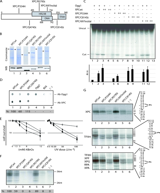
XPC is responsible for DNA damage sensing in nucleotide excision repair (NER). Mutations in XPC lead to a defect in NER and to xeroderma pigmentosum (XP-C). Here, we analyzed the biochemical properties behind mutations found within three patients: one amino acid substitution (P334H, XP1MI, and GM02096), one amino acid incorporation in a conserved domain (697insVal, XP8BE, and GM02249), and a stop mutation (R579St, XP67TMA, and GM14867). Using these mutants, we demonstrated that HR23B stabilizes XPC on DNA and protects it from degradation. XPC recruits the transcription/repair factor TFIIH and stimulates its XPB ATPase activity to initiate damaged DNA opening. In an effort to understand the severity of XP-C phenotypes, we also demonstrated that single mutations in XPC perturb other repair processes, such as base excision repair (e.g., the P334H mutation prevents the stimulation of Ogg1 glycosylase because it thwarts the interaction between XPC and Ogg1), thereby leading to a deeper understanding of the molecular repair defect of the XP-C patients.


Figures

FIG. 1.
FIG. 1.
DNA repair activities of the recombinant XPC-HR23B. (A) Diagram of XPC mutations and domains. (B) Purified XPC-HR23B complexes were subjected to sodium dodecyl sulfate-polyacrylamide gel electrophoresis and stained with Coomassie blue (upper panel) or detected by immunoblotting with a XPC antibody that recognizes the N-terminal region (lower panel). The asterisk corresponds to HR23B. (C) A DNA fragment containing a single 8-OH-Gua lesion was incubated with 60 fmol of Ogg1 and 100 (+) or 200 (++) fmol of purified XPCwt and XPC mutants, as indicated, for 30 min at 37°C. The Ogg1 activity was measured by densitometric scanning and is expressed in arbitrary units from at least three independent experiments. (D) Far Western analysis of Ogg1 binding capacities of XPC proteins was performed as described previously (12). Ogg1 and XPC levels were tested by probing either with anti-XPC or anti-Ogg1 polyclonal antibodies (lanes 1 to 4). Ogg1, bovine serum albumin (BSA), and buffer alone were used as negative controls (lanes 4 to 6). (E) Cell survival of GM02096 (XPC/P334H, closed rectangles), GM14867 (XPC/R579St, closed triangles), and wild-type fibroblasts (FB789, open circles) following either KBrO3 (left graph) or UV-C (right graph) treatments. (F) Portions (10 ng) of each of the purified XPC-HR23B complexes (lanes 1 to 6) were tested in an in vitro NER assay (lane 7, no XPC added). (G) KMnO4 footprint using 10 ng of XPC-HR23B (upper), plus 300 ng of TFIIH (middle), as well as all of the other NER factors, except XPF-ERCC1 (lower panel). Arrows indicate KMnO4 sensitive sites (3′ sense is denoted as +N, and 5′ sense is denoted as -N).
FIG. 2.
FIG. 2.
Recruitment of XPC and TFIIH at sites of UV damage. GM14867 fibroblasts were transiently transfected with pEGFP-XPC constructs and locally UV irradiated (100 J/m2). Cells were allowed to repair for 15 min at 37°C and immunoblotted with monoclonal anti-CPD (a, d, g, j, and s), polyclonal anti-full-length XPC (b and k), polyclonal anti-GFP (e, h, and t), or monoclonal anti-XPB (m and p). Panels n and q correspond to the direct GFP signal.
FIG. 3.
FIG. 3.
DNA-binding capacities of XPC proteins. (A) Purified XPC-HR23B complexes were incubated with a DNA fragment containing a cisplatin lesion immobilized on streptavidin beads (- columns), in the presence of TFIIH (IIH columns) or all of the dual incision factors (T columns). The values correspond to the quantification of the immunoblot bands of the XPC retained in the DNA, as a percentage of the protein input. (B) Amounts of either XPCwt and XPC/697insVal (upper panel) or XPCwt and XPC/P334H (lower panel), remaining on the immobilized damaged DNA after washing at 0.1, 0.2, and 0.3 M KCl. The graph represents the association between HR23B and XPCwt (⧫) or XPC/697insVal (formula image
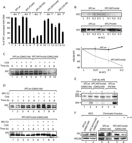
), when washed with 0.07 to 0.2 M KCl as indicated (lower graph). Error bars correspondent to the standard error of the mean of two independent experiments. (C) GM02184 (XPCwt) or GM02249 (XPC/697insVal) human lymphoblasts were incubated overtime with 0.1 mM CHX. Cell extracts were then analyzed by sodium dodecyl sulfate-polyacrylamide gel electrophoresis and immunoblotting with an XPC antibody. The asterisk corresponds to a nonspecific band. (D) GM02184 (XPCwt) or GM02249 (XPC/697insVal) human lymphoblasts were incubated during the indicated times with CHX either alone or in combination with MG132. Cell extracts were analyzed as described in panel C. The arrow indicates a probable degradation product (55). (E) ChIP followed by Western blot analysis of Ab-XPB immunoprecipitated samples from GM02184 (XPCwt), GM02249 (XPC/697insVal), and FB789 (XPCwt) cell lines fixed at t = 0 (no UV) or t = 15 min after UV irradiation (20 J/m2). A total of 400 μg of formaldehyde cross-linked protein extract was used per immunoprecipitation. (F) Portions (100 μg) of whole-cell extracts (WCE) or 40 μg of chromatin fraction extract from GM02184 or GM02249 cells were immunoblotted for the presence of XPC and HR23B. The asterisk corresponds to a nonspecific band.
FIG. 4.
FIG. 4.
Recruitment of XPC and XPA at sites of UV damage. (A) XPC deficient human primary fibroblasts (GM14867) were transiently transfected with either pEGFP-XPCwt or pEGFP-XPC/P334H and UV irradiated 24 h posttransfection. Cells were allowed to repair for the indicated times and immunoblotted with a monoclonal anti-XPA antibody (a, d, and g). The XPC signal corresponds to direct fluorescence from GFP (b, e, and h). Values correspond to increased fluorescent signal at UV spots in relation to the background. (B) ChIP followed by Western blot analysis (17) of Ab-XPB immunoprecipitated samples from FB789, GM02096 (XPC/P334H), and GM14867 (XPC/R579St) cell lines and fixed at t = 0, 15, and 30 min after UV irradiation. Portions (400 μg) of formaldehyde cross-linked extract were used per immunoprecipitation. (C) Portions (100 μg) of whole-cell extracts from XPC and XPB or p62 transfected Sf9 cells were immunoprecipitated with specific anti-p62 or anti-XPB antibodies (lanes 1 and 4, 10% of load; lane 2 and 5, XPC coimmunoprecipitated blotted with an antibody against XPC; lane 3, XPB; and lane 6, p62 immunoprecipitated).
FIG. 5.
FIG. 5.
Mutations in XPC impair the stimulation of TFIIH ATPase. (A) A portion (10 ng) of purified XPB was tested in an ATPase assay in the presence of 20 (+) or 50 (++) ng of p52 and of 20 (+) or 40 (++) ng of XPC proteins as indicated (lanes 1 to 12). Quantification of the release of the inorganic phosphate (Pi) and ATP was done by using a Bio-Imaging analyzer. (B) Portions (50 ng) of XPC-HR23B complexes were tested in an ATPase assay in the presence of 100 ng of TFIIH and 120 ng of DNA. Lane 6, ATP only. Graph corresponds to the quantification of two independent experiments. (C) Coimmunoprecipitation experiments between XPC and different truncated XPB proteins, using a specific monoclonal anti-XPB antibody.
FIG. 6.
FIG. 6.
(A) Scheme showing the XPC protein, indicating the mutations studied in the present study (boxes correspond to patient mutations), the interacting domains/regions identified thus far, and the role of some regions identified in the present study (the XPA binding region was described in reference 5). (B) First steps of the NER reaction. XPC, after UV irradiation, is stabilized by HR23B and recognizes and bends locally the damaged DNA; this bending precedes the recruitment of TFIIH through a double contact between XPC and the subunits p62 and XPB. This is followed by the release of HR23B concomitantly after. The ATPase of XPB is then regulated by XPC and p52. This results in a reorganization of the XPC/TFIIH/damaged DNA, which is then ready to recruit XPA and the other NER factors.

Similar articles

Cited by

References

    1. Aburatani, H., Y. Hippo, T. Ishida, R. Takashima, C. Matsuba, T. Kodama, M. Takao, A. Yasui, K. Yamamoto, and M. Asano. 1997. Cloning and characterization of mammalian 8-hydroxyguanine-specific DNA glycosylase/apurinic, apyrimidinic lyase, a functional mutM homologue. Cancer Res. 572151-2156. - PubMed
    1. Araujo, S. J., E. A. Nigg, and R. D. Wood. 2001. Strong functional interactions of TFIIH with XPC and XPG in human DNA nucleotide excision repair, without a preassembled repairosome. Mol. Cell. Biol. 212281-2291. - PMC - PubMed
    1. Bjoras, M., L. Luna, B. Johnsen, E. Hoff, T. Haug, T. Rognes, and E. Seeberg. 1997. Opposite base-dependent reactions of a human base excision repair enzyme on DNA containing 7,8-dihydro-8-oxoguanine and abasic sites. EMBO J. 166314-6322. - PMC - PubMed
    1. Bootsma, D., K. H. Kraemer, J. E. Cleaver, and J. H. J. Hoeijmakers. 2002. Nucleotide excision repair syndromes: xeroderma pigmentosum, Cockayne syndrome, and trichothiodystrophy, p. 211-237. In B. Vogelstein and K. W. Kinzler (ed.), The genetic basis of human cancer, 2nd ed. McGraw-Hill, New York, NY.
    1. Bunick, C. G., M. R. Miller, B. E. Fuller, E. Fanning, and W. J. Chazin. 2006. Biochemical and structural domain analysis of xeroderma pigmentosum complementation group C protein. Biochemistry 4514965-14979. - PMC - PubMed

Publication types