. 2008 Dec;28(23):7066-80.
doi: 10.1128/MCB.00244-08. Epub 2008 Sep 22.

Regulatory role of human AP-endonuclease (APE1/Ref-1) in YB-1-mediated activation of the multidrug resistance gene MDR1

Affiliations

Regulatory role of human AP-endonuclease (APE1/Ref-1) in YB-1-mediated activation of the multidrug resistance gene MDR1

Ranajoy Chattopadhyay et al. Mol Cell Biol. 2008 Dec.

Abstract

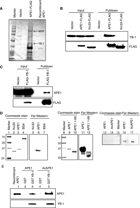
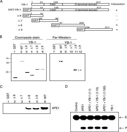
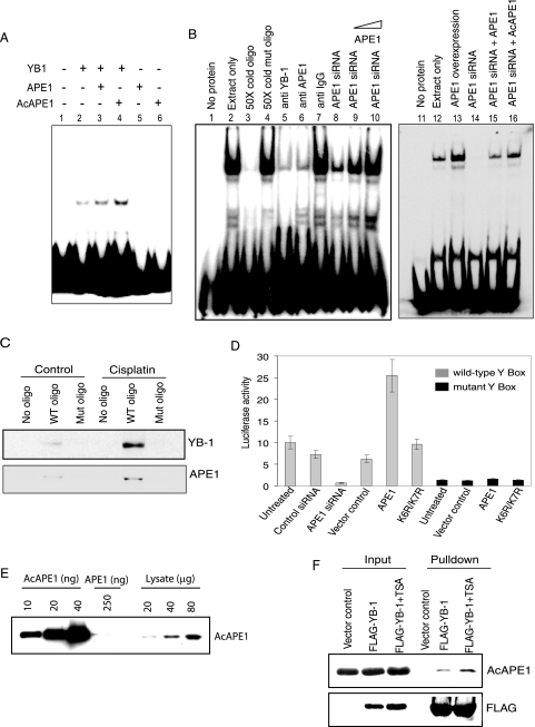
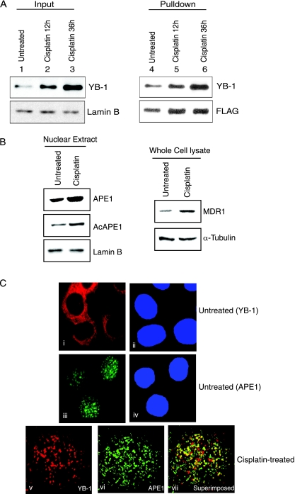
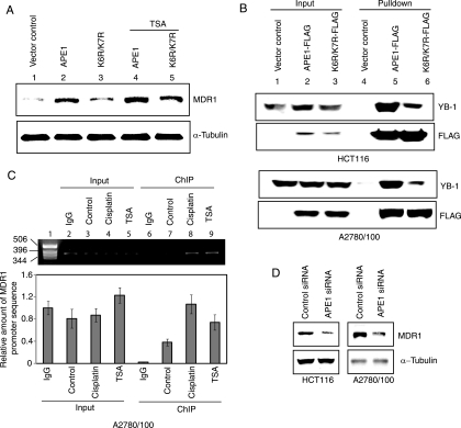
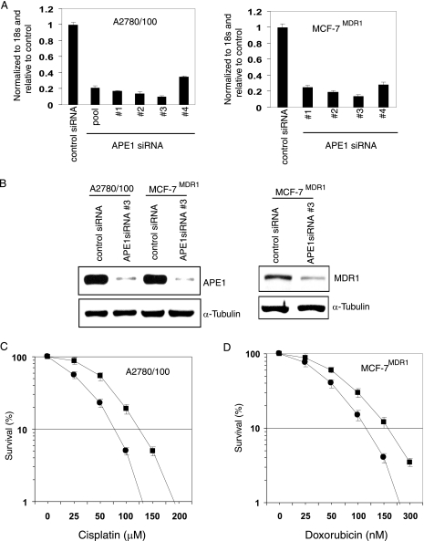
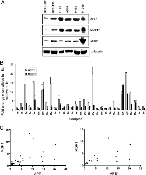
Human AP-endonuclease (APE1/Ref-1), a central enzyme involved in the repair of oxidative base damage and DNA strand breaks, has a second activity as a transcriptional regulator that binds to several trans-acting factors. APE1 overexpression is often observed in tumor cells and confers resistance to various anticancer drugs; its downregulation sensitizes tumor cells to such agents. Because the involvement of APE1 in repairing the DNA damage induced by many of these drugs is unlikely, drug resistance may be linked to APE1's transcriptional regulatory function. Here, we show that APE1, preferably in the acetylated form, stably interacts with Y-box-binding protein 1 (YB-1) and enhances its binding to the Y-box element, leading to the activation of the multidrug resistance gene MDR1. The enhanced MDR1 level due to the ectopic expression of wild-type APE1 but not of its nonacetylable mutant underscores the importance of APE1's acetylation in its coactivator function. APE1 downregulation sensitizes MDR1-overexpressing tumor cells to cisplatin or doxorubicin, showing APE1's critical role in YB-1-mediated gene expression and, thus, drug resistance in tumor cells. A systematic increase in both APE1 and MDR1 expression was observed in non-small-cell lung cancer tissue samples. Thus, our study has established the novel role of the acetylation-mediated transcriptional regulatory function of APE1, making it a potential target for the drug sensitization of tumor cells.


Figures

FIG. 1.
FIG. 1.
Stable interaction between YB-1 and APE1. (A) Identification of YB-1 as an APE1-associated protein. Extracts of HCT116 cells transfected with FLAG-tagged APE1 or empty vector were immunoprecipitated with FLAG antibody, and bound proteins were eluted with FLAG peptide, resolved by SDS-PAGE, and visualized with Coomassie stain. YB-1 and MVP (marked by arrow) were identified by matrix-assisted laser desorption ionization-time of flight analysis. (B, top) Extracts of HCT116 cells transfected with empty vector or WT APE1-FLAG or NΔ33APE1-FLAG were immunoprecipitated with FLAG antibody for Western analysis with YB-1 antibody. (Bottom) Western analysis of the same blot with FLAG antibody. (C) Western analysis of the FLAG-YB-1 immunoprecipitate with APE1 antibody (top) or with FLAG antibody (bottom). (D) Far-Western analysis with YB-1 antibody using the WT or NΔ33APE1 (left), the GST-tagged N-terminal 69 amino acid residues of APE1 (middle), and unmodified APE1 or AcAPE1 (right). Bovine serum albumin (BSA) was used as a control. (E) GST-tagged YB-1 was incubated separately with unmodified APE1 (lane 3) or in vitro-acetylated APE1 (lane 5). Bound proteins were eluted and immunoblotted with APE1 antibody. (Bottom) Western analysis of the same blot with YB-1 antibody.
FIG. 2.
FIG. 2.
Mapping of the APE1-interacting region in YB-1 by far-Western and GST pull-down analyses. (A) Schematic representation of the GST-YB1 deletion mutants. CSD, cold shock domain. (B) Far-Western analysis. (Right) APE1 antibody was used to test different GST-YB-1 deletion mutants. (Left) Coomassie staining of YB-1 deletion mutants. (C) GST pull-down assay. Recombinant APE1 was incubated with glutathione-agarose affinity beads containing different GST-YB-1 deletion mutants (Δ6 to Δ9, as indicated), and the eluted proteins were immunoblotted with APE1 antibody. (D) Effect of YB-1 on APE1's AP-endonuclease activity. APE1 (50 pM) was incubated for 10 min with the tetrahydrofuran-containing 32P-labeled duplex in the presence of increasing amounts of YB-1 (0.05 to 5 nM), and the reaction products were analyzed in a 20% urea-polyacrylamide gel as described previously (7). S, substrate; P, product.
FIG. 3.
FIG. 3.
APE1 enhances YB-1 binding to the Y-box sequence. (A) EMSA of YB-1 (10 ng) with or without added APE1 (20 ng) or AcAPE1 (20 ng) using 32P-labeled duplex oligonucleotides containing the WT Y-box sequence. Lanes 5 and 6, APE1 or AcAPE1 alone. (B) APE1 enhances binding of HCT116 NE to 5′ 32P-labeled Y-box-containing oligonucleotide (oligo). Lane 1, free probe. Lanes 2 to 16, EMSA with NE (3 μg) alone or in the presence of 50-fold unlabeled WT or mutant oligonucleotide (lanes 2 to 4); NE preincubated with anti-YB-1, anti-APE1, and control rabbit IgG (lanes 5 to 7); NE from APE1 siRNA-treated cells alone or supplemented with 100 ng or 500 ng recombinant APE1 (lanes 8 to 10); and NE from cells expressing ectopic APE1 (lane 13) or APE1 siRNA-treated cells alone (right, lane 14) or supplemented with 500 ng of APE1 (lane 15) or recombinant AcAPE1 (lane 16). (C) Western analysis of eluted proteins bound to 5′ biotin-labeled WT or mutant Y-box-containing oligonucleotide with YB-1 (top) or APE1 (bottom) antibody. (D) HCT116 cells were cotransfected with a MDR1 promoter-driven luciferase reporter plasmid containing either the WT (gray) or mutant (black) Y box and expression plasmids for APE1 (WT or the K6R/K7R mutant) or equivalent amounts of empty vector. Where indicated, cells were transfected with APE1-specific siRNA or control siRNA (80 nM) 24 h before transfection with the reporter plasmid. Luciferase activity was normalized with coexpressed β-galactosidase. The bar graph shows the averages ± standard deviations for three independent experiments performed in duplicate. (E) Western analysis of recombinant AcAPE1 and HCT116 cell extracts with AcAPE1-specific antibody. (F) HCT116 cells were transfected with FLAG-YB-1 and treated 24 h later with TSA (100 ng/ml) for 12 h. Immunoprecipitates of the cell extracts with FLAG antibody were analyzed by Western blotting with AcAPE1 (top) or FLAG antibody (bottom).
FIG. 4.
FIG. 4.
Enhanced interaction between APE1 and YB-1 after cisplatin treatment. (A) HCT116 cells transfected with APE1-FLAG were treated with 40 μg/ml cisplatin for 12 h (lanes 2 and 5) and 36 h (lanes 3 and 6). NEs were immunoprecipitated with FLAG antibody beads, followed by Western analysis using YB-1 antibody (top right) or FLAG antibody (bottom right). (Left) Western analysis for YB-1 in input nuclear extract (lamin B as the loading control). (B) Western analysis of APE1, AcAPE1, and MDR1 in nuclear (left) and whole-cell (right) extracts after cisplatin treatment for 12 h. Lamin B (bottom left) or α-tubulin (bottom right) was used as a loading control. (C) Colocalization of APE1 and YB-1 after cisplatin treatment. HCT116 cells were immunostained with rabbit YB-1 (i and v) and mouse APE1 antibody (iii and vi) followed by goat anti-rabbit Texas Red-labeled and goat anti-mouse fluorescein isothiocyanate-labeled secondary antibody. vii, superimposition of images v and vi. DAPI (4′,6′-diamidino-2-phenylindole) was used for nuclear staining (ii and iv).
FIG. 5.
FIG. 5.
APE1 acetylation-dependent enhancement of MDR1 expression. (A) A2780/100 cells were transfected with expression plasmids for WT (lanes 2 and 4) or K6R/K7R (lanes 3 and 5) APE1. After 24 h, one set of cells was treated with 100 ng/ml TSA (lanes 4 and 5) for 12 h, and the MDR1 level was analyzed by Western blotting with MDR1 antibody. (B, top) Extracts of HCT116 and A2780/100 cells transfected with FLAG-tagged WT APE1 or K6R/K7R APE1 were immunoprecipitated with FLAG antibody beads and analyzed by Western blotting using YB-1 antibody. (Bottom) Western analysis of the same blot with FLAG antibody. (C) ChIP assay for in vivo association of APE1 with the MDR1 promoter. (Top) PCR analysis of immunoprecipitated MDR1 promoter sequence. Lane 1, DNA molecular weight marker; lanes 2 to 5, MDR1 promoter sequence in control IgG, untreated control, and cisplatin- and TSA-treated input chromatin, respectively; lanes 7 to 9, immunoprecipitated MDR1 promoter sequence with APE1 antibody from control and cisplatin- and TSA-treated cell extracts, respectively; lane 6, immunoprecipitated MDR1 promoter with control IgG. (Bottom) Quantitation of immunoprecipitated MDR1 promoter DNA by real-time PCR analysis as described in Materials and Methods. (D) HCT116 and A2780/100 cells were transfected with 80 nM APE1 siRNA oligonucleotide. The MDR1 level (top) was measured by Western analysis at 48 h after transfection (α-tubulin was used as the loading control).
FIG. 6.
FIG. 6.
APE1 downregulation sensitizes drug-resistant cells. (A) Real-time RT-PCR analysis of APE1 mRNA from A2780/100 (left) and MCF-7MDR1 (right) cells at 48 h after transfection with 80 nM of APE1-specific or control duplex siRNAs. (B, left) Western analysis of cell extracts with APE1 (top) or α-tubulin (bottom) antibody. (Right) Western analysis of MDR1 levels in MCF-7 MDR1 cells at 48 h after transfection with 80 nM APE1 siRNA oligonucleotide. (C and D) Survival assay of A2780/100 and MCF-7MDR1 cells transfected with either control (▪) or APE1 (•) siRNA; 48 h after transfection, ∼300 cells were plated into 60-mm dishes and treated with increasing concentrations of cisplatin (0 to 200 μM) or doxorubicin (0 to 300 nM). Cells were maintained for 10 days with or without drug treatment, and the colonies were counted using crystal violet. One hundred percent corresponds to the colony numbers in the control without drug treatment. Other details are given in Materials and Methods.
FIG. 7.
FIG. 7.
MDR1 expression is correlated with APE1 levels in normal lung and NSCLC tissues. (A) Western analysis of APE1, AcAPE1, and MDR1 levels in cell extracts of various lung cancer cell lines. α-Tubulin was used as the loading control. (B) Real-time RT-PCR analysis of APE1 and MDR1 mRNA expression levels in 15 NSCLC specimens and proximal normal lung tissues as described in Materials and Methods. The level of APE1 mRNA was expressed relative to the level in normal lung tissue of patient 1n,which was normalized to 1. n, normal lung tissues; c, NSCLC tissues. The bar graph shows the averages ± standard deviations for three independent experiments performed in duplicates. (C) Correlation between APE1 and MDR1 expression levels in both normal and NSCLC samples together (left) (r = 0.581; P = 0.0005) and in NSCLC samples (right) (r = 0.558; P = 0.03).

Similar articles

Cited by

References

    1. Bargou, R. C., K. Jurchott, C. Wagener, S. Bergmann, S. Metzner, K. Bommert, M. Y. Mapara, K. J. Winzer, M. Dietel, B. Dorken, and H. D. Royer. 1997. Nuclear localization and increased levels of transcription factor YB-1 in primary human breast cancers are associated with intrinsic MDR1 gene expression. Nat. Med. 3447-450. - PubMed
    1. Bhakat, K. K., T. Izumi, S. H. Yang, T. K. Hazra, and S. Mitra. 2003. Role of acetylated human AP-endonuclease (APE1/Ref-1) in regulation of the parathyroid hormone gene. EMBO J. 226299-6309. - PMC - PubMed
    1. Bhakat, K. K., S. K. Mokkapati, I. Boldogh, T. K. Hazra, and S. Mitra. 2006. Acetylation of human 8-oxoguanine-DNA glycosylase by p300 and its role in 8-oxoguanine repair in vivo. Mol. Cell. Biol. 261654-1665. - PMC - PubMed
    1. Bobola, M. S., L. S. Finn, R. G. Ellenbogen, J. R. Geyer, M. S. Berger, J. M. Braga, E. H. Meade, M. E. Gross, and J. R. Silber. 2005. Apurinic/apyrimidinic endonuclease activity is associated with response to radiation and chemotherapy in medulloblastoma and primitive neuroectodermal tumors. Clin. Cancer Res. 117405-7414. - PubMed
    1. Boeckman, H. J., K. S. Trego, and J. J. Turchi. 2005. Cisplatin sensitizes cancer cells to ionizing radiation via inhibition of nonhomologous end joining. Mol. Cancer Res. 3277-285. - PMC - PubMed

Publication types

MeSH terms

Substances