. 2008 Nov 21;283(47):32198-208.
doi: 10.1074/jbc.M803446200. Epub 2008 Sep 22.

G9a and HP1 couple histone and DNA methylation to TNFalpha transcription silencing during endotoxin tolerance

Affiliations

G9a and HP1 couple histone and DNA methylation to TNFalpha transcription silencing during endotoxin tolerance

Mohamed El Gazzar et al. J Biol Chem. .

Abstract

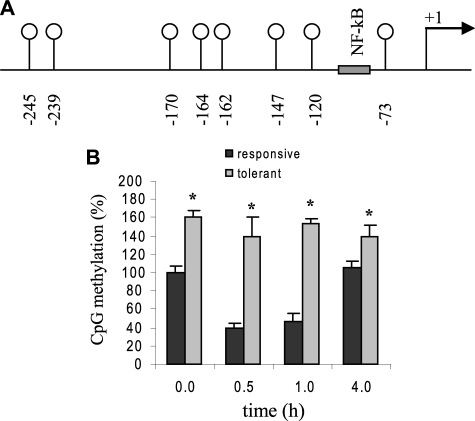
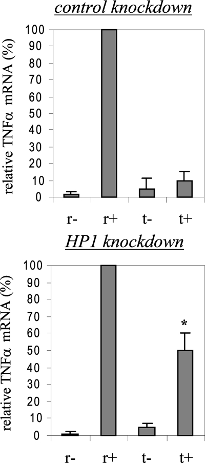
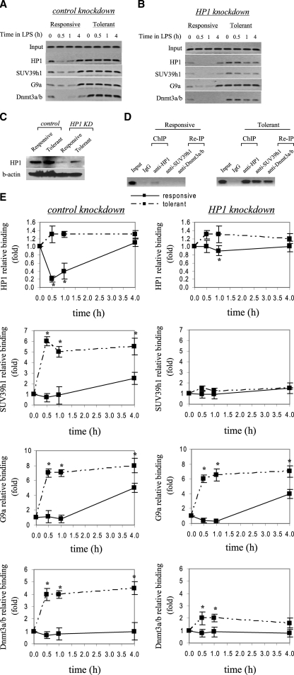
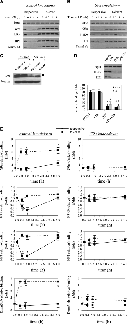
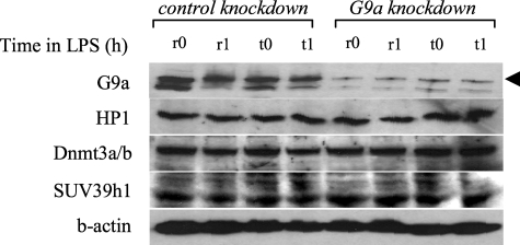
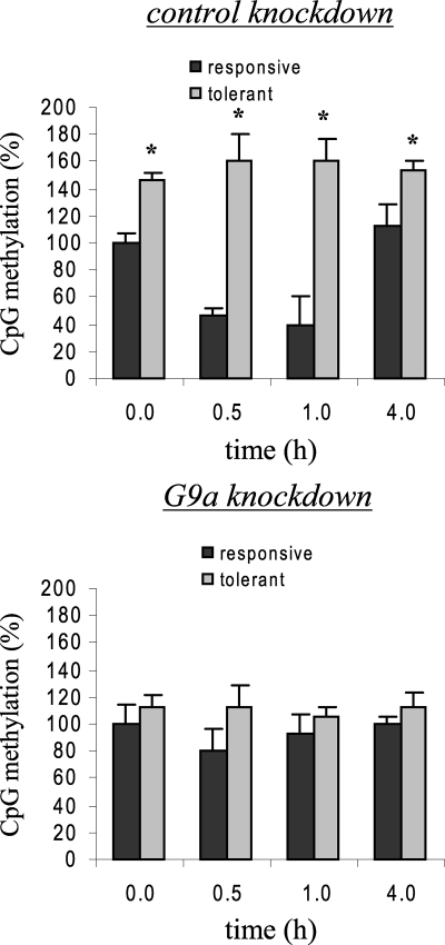
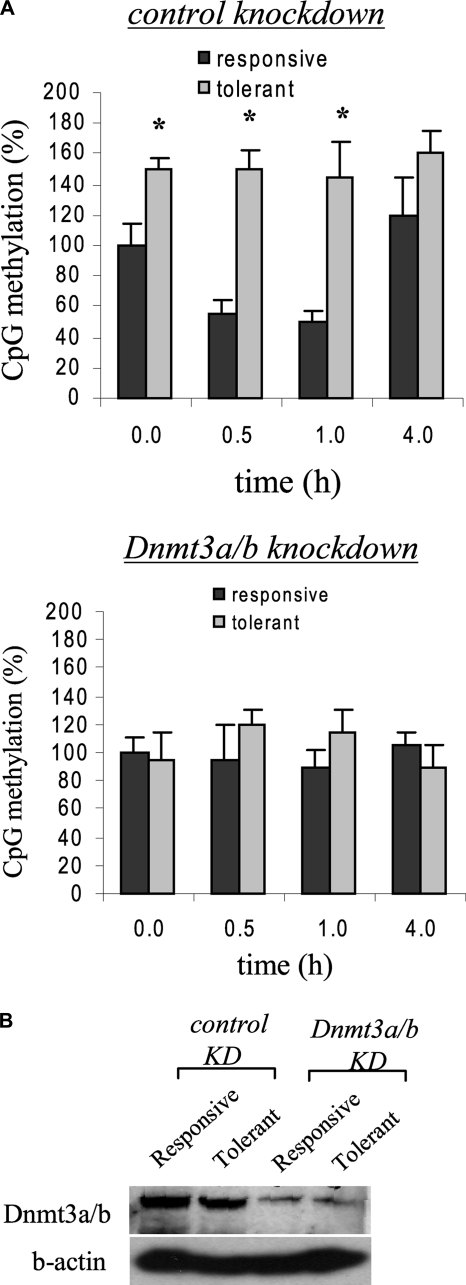
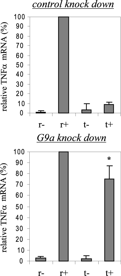
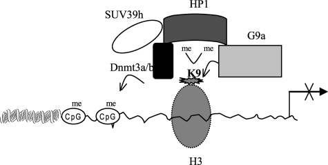
TNFalpha gene expression is silenced in the endotoxin tolerant phenotype that develops in blood leukocytes after the initial activation phase of severe systemic inflammation or sepsis. The silencing phase can be mimicked in vitro by LPS stimulation. We reported that the TNFalpha transcription is disrupted in endotoxin tolerant THP-1 human promonocyte due to changes in transcription factor binding and enrichment with histone H3 dimethylated on lysine 9 (H3K9). Here we show that the TNFalpha promoter is hypermethylated during endotoxin tolerance and that H3K9 methylation and DNA methylation interact to silence TNFalpha expression. Chromatin immunoprecipitation and RNA interference analysis demonstrated that, in tolerant cells, TNFalpha promoter is bound by the H3K9 histone methyltransferase G9a which dimethylates H3K9 and creates a platform for HP1 binding, leading to the recruitment of the DNA methyltransferase Dnmt3a/b and an increase in promoter CpG methylation. Knockdown of HP1 resulted in a decreased Dnmt3a/b binding, sustained G9a binding, and a modest increase in TNFalpha transcription, but had no effect on H3K9 dimethylation. In contrast, G9a knockdown-disrupted promoter silencing and restored TNFalpha transcription in tolerant cells. This correlated with a near loss of H3K9 dimethylation, a significant decrease in HP1 and Dnmt3a/b binding and promoter CpG methylation. Our results demonstrate a central role for G9a in this process and suggest that histone methylation and DNA methylation cooperatively interact via HP1 to silence TNFalpha expression during endotoxin tolerance and may have implication for proinflammatory gene silencing associated with severe systemic inflammation.


Figures

FIGURE 1.
FIGURE 1.
TNFα promoter is hypermethylated in endotoxin-tolerant THP-1 cells. A, a schematic diagram representing the proximal promoter region of the human TNFα gene analyzed by bisulfite sequencing. The eight CpG dinucleotides located in this region are represented by vertical lines with open circles, and their locations relative to the transcription start site are indicated. B, bisulfite sequencing results. Endotoxin tolerance was induced by pretreatment with 1 μg/ml LPS for 16 h. Endotoxin responsive and tolerant cells were then stimulated with 1 μg/ml LPS for the indicated times. Genomic DNA was extracted and subjected to sodium bisulfite conversion, followed by PCR amplification and sequencing. The total numbers of methylated CpGs in responsive untreated cells (0 h) was set at 100%, and all other values were calculated relative to this value. The data represent the mean ± S.E. of at least three independent experiments. *, significant difference (p < 0.05) between responsive versus tolerant cells.
FIGURE 2.
FIGURE 2.
HP1 gene silencing modestly restores TNFα mRNA level in tolerant cells. THP-1 cells were transfected with nonspecific or HP1-specific siRNA and then left unstimulated or stimulated with 1 μg/ml LPS for 36 h. Responsive (r) and tolerant (t) were washed and then left unstimulated or stimulated with 1 μg/ml LPS for 3 h. Total RNA was extracted and analyzed for TNFα mRNA expression by real-time PCR. Sample data were normalized to GAPDH data and are presented as percent change relative to mRNA level in LPS-responsive (r+) cells (set as 100%). Data are the mean ± S.E. of at least three independent experiments. *, significant difference (p < 0.05), t+ versus t-.
FIGURE 3.
FIGURE 3.
HP1 gene silencing markedly reduces SUV39h1, Dnmt3a/b, but not G9a binding to the TNFα promoter. THP-1 cells were transfected with control or HP1-specific siRNA and then left unstimulated or stimulated with 1 μg/ml LPS for 36 h. Cells were then washed and left unstimulated or stimulated with 1 μg/ml LPS for the indicated times. ChIP assay was performed on cross-linked chromatin, immunoprecipitated with antibodies specific to HP1, SUV39h1, G9a, and Dnmt3a/b. The relative enrichment of TNFα promoter sequences in the immunoprecipitated DNA was analyzed by semiquantitative PCR (A and B) and real-time PCR (E) using primers that amplify the proximal promoter region. In A and B, representative results are shown. In E, data were normalized to input DNA and are presented as fold change relative to unstimulated cells (0 h) (set as 1-fold). Data are the mean ± S.E. of at least three independent experiments. *, significant difference (p < 0.05). C, HP1 protein expression in the nucleus. THP-1 cells were transfected with control or HP1-specific siRNA and left unstimulated or stimulated with 1 μg/ml LPS for 36 h. Responsive (unstimulated) and tolerant (stimulated) cells were then washed and stimulated for 1 h with 1 μg/ml LPS, and nuclear extract was prepared as described under “Experimental Procedures.” Blotted proteins were probed with HP1 Ab. Blots were stripped and reprobed with control Ab. D, ChIP reimmunoprecipitation showing the recruitment of SUV39h1 and Dnmt3a/b to the TNFα promoter by HP1. Chromatin was first immunoprecipitated with HP1 Ab (ChIP). Immunoprecipitates were then eluted and reimmunoprecipitated (Re-IP) with SUV39h1 or Dnmt3a/b Ab.
FIGURE 4.
FIGURE 4.
G9a gene silencing significantly decreases H3K9 dimethylation and promoter binding by HP1 and Dnmt3a/b. Cells were transfected with control or G9a-specific siRNA and then left unstimulated or stimulated with 1 μg/ml LPS for 36 h. Cells were then washed and left unstimulated or stimulated with 1 μg/ml LPS for the indicated times. ChIP assay was performed on cross-linked chromatin, immunoprecipitated with antibodies specific to HP1, G9a, dimethyl H3K9, and Dnmt3a/b. The relative enrichment of TNFα promoter sequences in the immunoprecipitated DNA was analyzed by semiquantitative PCR (A and B) and real-time PCR (E), using primers that amplify the proximal promoter region. In A and B, representative results are shown. In E, data were normalized to input DNA and are presented as fold change relative to unstimulated cells (0 h) (set as 1-fold). Data are the mean ± S.E. of at least three independent experiments. *, significant difference (p < 0.05). C, G9a protein expression in the nucleus. THP-1 cells were transfected with control or G9a-specific siRNA and left unstimulated or stimulated with 1 μg/ml LPS for 36 h (to induce tolerance). Responsive (unstimulated) and tolerant (stimulated) cells were then washed and stimulated for 1 h with 1 μg/ml LPS, and nuclear extract was prepared as described under “Experimental Procedures.” Blotted proteins were probed with G9a Ab. Blots were stripped and reprobed with control Ab. D, inhibiting H3K9 dimethylation prevents HP1 binding. LPS-tolerant cells were incubated for 6 h with 6μm of the G9a specific inhibitor BIX-012194. LPS was added for the last 1 h. Cells were harvested, and ChIP was performed with dimethyl H3K9 or HP1 Ab. Enrichment of TNFα promoter sequences in the immunoprecipitated DNA was assessed by semiquantitative PCR (upper panel) and real-time PCR (lower panel).
FIGURE 5.
FIGURE 5.
Western blot analysis of whole cell extract after G9a knockdown. Cells were transfected with control or G9a-specific siRNA and left unstimulated or stimulated with 1 μg/ml LPS for 36 h (to induce tolerance). Responsive (unstimulated) and tolerant (stimulated) cells were then washed and stimulated for 0.5 h and 1 h with 1 μg/ml LPS. Whole cell lysates were prepared and analyzed for the expression of G9a, HP1, Dnmt3a/b, and SUV39h1. Blots were first probed with G9a Ab, stripped, and reprobed with HP1, Dnmt3a/b, and SUV39h1 Abs.
FIGURE 6.
FIGURE 6.
Loss of G9a binding prevents TNFα promoter CpG hypermethylation in tolerant cells. Cells were transfected with control or G9a-specific siRNA and then left unstimulated or stimulated with 1 μg/ml LPS for 36 h. Endotoxin-responsive and -tolerant cells were then stimulated with 1 μg/ml LPS for the indicated times. Genomic DNA was extracted and subjected to sodium bisulfite conversion, followed by PCR amplification and sequencing. The total numbers of methylated CpGs in responsive untreated cells (0 h) was set at 100%, and all other values were calculated relative to this value. The data represent the mean ± S.E. of at least three independent experiments. *, significant difference (p < 0.05) between responsive versus tolerant cells. R, responsive; t, tolerant.
FIGURE 7.
FIGURE 7.
Dnmt3a is responsible for TNFα promoter methylation. A, cells were transfected with control or Dnmt3a/b-specific siRNA and left unstimulated or stimulated with 1 μg/ml LPS for 36 h (to induce tolerance). Responsive (unstimulated) and tolerant (stimulated) cells were then washed and stimulated with 1 μg/ml LPS for the indicated times. Genomic DNA was extracted and analyzed by bisulfite sequencing. The total number of methylated CpGs in responsive untreated cells (0 h) was set at 100%, and all other values were calculated relative to this value. The data represent the mean ± S.E. of at least three independent experiments. *, significant difference (p < 0.05) between responsive versus tolerant cells. B, Dnmt3a/b protein expression in the nucleus after the knockdown. Cells were transfected with Dnmt3a/b as described in A, and nuclear extract was prepared at 1 h after LPS stimulation and analyzed for Dnmt3a/b expression by Western blot.
FIGURE 8.
FIGURE 8.
Loss of G9a binding restores TNFα mRNA expression in tolerant cells. THP-1 cells were transfected with nonspecific or G9a-specific siRNA and then left unstimulated or stimulated with 1 μg/ml LPS for 36 h. Responsive (r) and tolerant (t) were washed and then left unstimulated or stimulated with 1 μg/ml LPS for 3 h. Total RNA was extracted and analyzed for TNFα mRNA expression by real-time PCR. Sample data were normalized to GAPDH data and are presented as percent change relative to mRNA level in LPS-responsive (r+) cells (set as 100%). Data are the mean ± S.E. of at least three independent experiments. *, significant difference (p < 0.05), t+ versus t-.
FIGURE 9.
FIGURE 9.
A model depicting a central role for G9a in proinflammatory gene silencing during endotoxin tolerance. G9a is first recruited to euchromatic regions, likely via a transcriptional repressor, where it dimethylates H3K9. The HP1 adaptor then binds to dimethylated H3K9 and recruits Dnmt3a/b to enhance cytosine methylation, resulting in transformation of euchromatin to silent heterochromatin and transcription silencing. In addition, HP1 may recruit SUV39h, which can trimethylate H3K9 on adjacent histone to reinforce the silent heterochromatin state.

References

    1. Brenner, C., and Fuks, F. (2007) Dev. Cell 12 843–844 - PubMed
    1. Dormann, H. L., Tseng, B. S., Allis, C. D., Funabiki, H., and Fischle, W. (2006) Cell Cycle 5 2842–2851 - PubMed
    1. Hiragami, K., and Festenstein, R. (2005) Cell Mol. Life Sci. 62 2711–2726 - PMC - PubMed
    1. Craig, J. M. (2005) Bioessays 27 17–28 - PubMed
    1. Chin, H. G., Esteve, P. O., Pradhan, M., Benner, J., Patnaik, D., Carey, M. F., and Pradhan, S. (2007) Nucleic Acids Res. 35 7313–7323 - PMC - PubMed

Publication types

LinkOut - more resources