. 2008 Sep 22;182(6):1185-200.
doi: 10.1083/jcb.200803045.

NCAM induces CaMKIIalpha-mediated RPTPalpha phosphorylation to enhance its catalytic activity and neurite outgrowth

Affiliations

NCAM induces CaMKIIalpha-mediated RPTPalpha phosphorylation to enhance its catalytic activity and neurite outgrowth

Vsevolod Bodrikov et al. J Cell Biol. .

Abstract

Receptor protein tyrosine phosphatase alpha (RPTPalpha) phosphatase activity is required for intracellular signaling cascades that are activated in motile cells and growing neurites. Little is known, however, about mechanisms that coordinate RPTPalpha activity with cell behavior. We show that clustering of neural cell adhesion molecule (NCAM) at the cell surface is coupled to an increase in serine phosphorylation and phosphatase activity of RPTPalpha. NCAM associates with T- and L-type voltage-dependent Ca(2+) channels, and NCAM clustering at the cell surface results in Ca(2+) influx via these channels and activation of NCAM-associated calmodulin-dependent protein kinase IIalpha (CaMKIIalpha). Clustering of NCAM promotes its redistribution to lipid rafts and the formation of a NCAM-RPTPalpha-CaMKIIalpha complex, resulting in serine phosphorylation of RPTPalpha by CaMKIIalpha. Overexpression of RPTPalpha with mutated Ser180 and Ser204 interferes with NCAM-induced neurite outgrowth, which indicates that neurite extension depends on NCAM-induced up-regulation of RPTPalpha activity. Thus, we reveal a novel function for a cell adhesion molecule in coordination of cell behavior with intracellular phosphatase activity.


Figures

Figure 1.
Figure 1.
NCAM140 clustering induces phosphorylation of RPTPα on Ser180 and Ser204 and enhances the phosphatase activity of RPTPα. (A) Graph shows phosphatase activity of RPTPα from NCAM+/+ and NCAM−/− brain lysates. (B–D) RPTPα immunoprecipitates were probed by Western blotting with antibodies against RPTPα and phospho serines (B and C) or subjected to serine phosphorylation estimation by the alkaline hydrolysis (D). Note that similar levels of RPTPα were immunoprecipitated from NCAM+/+ and NCAM−/− brain lysates (B). Mock immunoprecipitation with nonspecific IgG served as a control. Phosphatase activity and serine phosphorylation of RPTPα are reduced in NCAM−/− brains. Graph in C shows quantitation of the blots in B with optical density for NCAM+/+ brains set to 100%. In A and D, mean values of phosphatase activity (A) or phosphate released by alkaline (D) in RPTPα immunoprecipitates from NCAM+/+ brains were set to 100%. (E) RPTPα-negative fibroblasts transfected with RPTPαWT alone or cotransfected with NCAM140 and RPTPαWT, RPTPαS180A, RPTPαS204A, or RPTPαS180/204A were treated with NCAM polyclonal antibodies or nonspecific rabbit IgG. Note that similar levels of NCAM140 were expressed in cells (lysates) and that similar levels of RPTPα were immunoprecipitated from the lysates (IP: HA-tag). Mutation of Ser180 and/or Ser204 does not influence coimmunoprecipitation of NCAM140 with RPTPα. Immunoprecipitated RPTPα was then subjected to the serine phosphorylation estimation by the alkaline hydrolysis (top) and phosphatase activity analysis (bottom). Application of NCAM antibodies but not IgG increased serine phosphorylation and phosphatase activity of RPTPαWT in NCAM140–RPTPαWT–cotransfected cells. Phosphorylation and activation of RPTPα mutants were inhibited. Mean values of phosphate released by alkaline (top) or phosphatase activity (bottom) in RPTPα immunoprecipitates from IgG-treated NCAM140–RPTPαWT–cotransfected cells were set to 100%. Mean values ± SEM are shown (A and D, n ≥ 8; C, n = 6; E, n ≥ 6). *, P < 0.05 (paired t test).
Figure 2.
Figure 2.
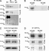
PKCδ associates with NCAM and RPTPα but its activity is not regulated by NCAM. (A and B) PKCδ (A) or NCAM (B) immunoprecipitates (IP) from NCAM+/+ brain lysates were analyzed by Western blotting as indicated. Mock immunoprecipitation with nonspecific IgG (A) or from NCAM−/− brain lysates (B) served as a control. RPTPα and NCAM120 coimmunoprecipitate with PKCδ, and PKCδ coimmunoprecipitates with NCAM. BH, brain homogenate. (C and D) NCAM+/+ and NCAM−/− brain homogenates (C) and RPTPα immunoprecipitates from NCAM+/+ and NCAM−/− brain lysates (D) were probed by Western blotting with antibodies against total PKCδ and active Thr505-phosphorylated PKCδ. Note the similar loading (GAPDH labeling in C) and immunoprecipitation efficiency (RPTPα labeling in D). Total levels of active PKCδ and levels of active PKCδ associated with RPTPα are not changed in NCAM−/− brains.
Figure 3.
Figure 3.
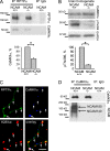
NCAM promotes CaMKIIα activation and RPTPα–CaMKIIα complex formation. (A) RPTPα immunoprecipitates (IP) from NCAM+/+ and NCAM−/− brain lysates were probed by Western blotting with antibodies against CaMKIIα. Note that similar levels of RPTPα were immunoprecipitated. Mock immunoprecipitation with nonspecific IgG served as control. Coimmunoprecipitation of CaMKIIα with RPTPα is reduced in NCAM−/− brain lysates. (B) NCAM+/+ and NCAM−/− brain homogenates were probed by Western blotting with antibodies against total and active Thr286-phosphorylated CaMKIIα and GAPDH (loading control). The levels of active CaMKIIα were reduced in NCAM−/− brain homogenates. Graphs show quantitation of the blots (mean ± SEM, n = 6 for A and B) with optical density for NCAM+/+ probes set to 100%. *, P < 0.05, paired t test. (C) NCAM was clustered at the cell surface of cultured hippocampal neurons by NCAM antibodies (H28live). Neurons were then fixed and colabeled with antibodies against RPTPα and CaMKIIα. A high-magnification image of a neurite is shown. NCAM clusters overlap with accumulations of RPTPα and CaMKIIα (arrows). Bar, 5 μm. (D) CaMKIIα immunoprecipitates from NCAM+/+ and NCAM−/− brain lysates were probed by Western blotting with antibodies against NCAM. NCAM140 and NCAM180 coimmunoprecipitated with CaMKIIα. Mock immunoprecipitation with nonspecific IgG served as a control.
Figure 4.
Figure 4.
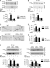
RPTPα is phosphorylated by CaMKIIα on Ser180 and Ser204. (A) Recombinant RPTPα-ID-GST or GST coupled to beads were incubated with CaMKIIα. The gel stained with Coomassie blue shows that approximately equal amounts of RPTPα-ID-GST and GST were bound to beads. The beads, treated and nontreated with CaMKIIα, were then subjected to the analysis of serine phosphorylation by alkaline hydrolysis (bottom left) or phosphatase activity (bottom right). Values for GST were subtracted as background, and values of serine phosphorylation (bottom left) or phosphatase activity (bottom right) for RPTPα-ID-GST not treated with CaMKIIα were set to 100%. (B) RPTPα from NCAM−/− brain lysates was incubated with CaMKIIα in the presence or absence of KN62 and Bis1. RPTPα was then subjected to the analysis of serine phosphorylation by alkaline hydrolysis. Values of phosphate released by alkaline from RPTPα not treated with CaMKIIα were set to 100%. (C) RPTPα immunoprecipitated from lysates of RPTPα-negative fibroblasts transfected with RPTPαWT, RPTPαS180A, RPTPαS204A, or RPTPαS180/204A was incubated with CaMKIIα. The immunoprecipitates, treated and untreated with CaMKIIα, were then subjected to the analysis of serine phosphorylation by alkaline hydrolysis (left) or phosphatase activity (right). Mean values of serine phosphorylation (left) or phosphatase activity (right) of RPTPαWT not treated with CaMKIIα were set to 100%. Mean values ± SEM are shown (A, n = 6; B, n = 11; C, n = 9). *, P < 0.05 (paired t test).
Figure 5.
Figure 5.
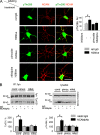
CaMKIIα activity is required for NCAM140-induced fyn activation. (A and B) CHO cells were cotransfected with NCAM140 and RPTPαWT and treated with nonspecific rabbit IgG or NCAM polyclonal antibodies in the absence or presence of KN62. RPTPα and fyn immunoprecipitates from the lysates of these cells were then probed with phosphoserine antibodies (A) or antibodies against activated Tyr531-dephosphorylated fyn (B) by Western blotting. Note that similar levels of RPTPα and fyn were immunoprecipitated in all groups. KN62 inhibited serine phosphorylation of RPTPα and activation of fyn in response to NCAM antibodies. Graphs show quantitation of the blots (mean ± SEM), with the levels in cells treated with nonspecific IgG without KN62 set to 100%. (C) Representative images of neurites of cultured hippocampal neurons treated with nonspecific rat IgG or NCAM monoclonal antibodies (H28live) in the presence or absence of KN62 and labeled with antibodies against activated Tyr416-phosphorylated fyn by indirect immunofluorescence. Inverted immunofluorescence images are shown to accentuate the differences in labeling intensities. Note the higher levels of activated fyn along neurites of NCAM antibody-treated neurons and the inhibition of this effect by KN62. Bar, 10 μm. (D and E) Graphs show mean levels ± SEM in arbitrary units of active Tyr416-phosphorylated (D) or Tyr531-dephosphorylated (E) fyn along neurites of hippocampal neurons treated with nonspecific rat IgG or NCAM monoclonal antibodies (H28live, D), or Fc or NCAM-Fc (E) in the absence or presence of KN62. n > 90 neurites from 45 neurons from 3 coverslips analyzed in each group (for D and E). (F) Cultured cortical neurons were treated with nonspecific rabbit IgG or NCAM polyclonal antibodies in the absence or presence of BisI and KN62. Lysates of these cells and fyn immunoprecipitates from the lysates were then probed with the indicated antibodies by Western blotting. Note that KN62 but not BisI inhibited fyn and CaMKIIα activation in response to NCAM antibodies. Graphs show quantitation of the blots (mean ± SEM) with the levels in cells treated, with nonspecific IgG without inhibitors set to 100%. *, P < 0.05 (paired t test).
Figure 6.
Figure 6.
Association of NCAM140 with lipid rafts is required for CaMKIIα activation. (A, left) Representative images of neurites of cultured hippocampal neurons treated with nonspecific rabbit IgG or NCAM polyclonal antibodies, extracted in cold 1% Triton X-100, and labeled with antibodies against activated Thr286-phosphorylated CaMKIIα and PI(4,5)P2 by indirect immunofluorescence. Note the higher levels of activated CaMKIIα and the higher degree of colocalization of activated CaMKIIα with PI(4,5)P2 along neurites of NCAM antibody-treated neurons. Arrows show clusters of activated CaMKIIα colocalizing with PI(4,5)P2 accumulations. Bar, 10 μm. (right) Graphs show mean levels ± SEM in arbitrary units (AU) of active Thr286-phosphorylated CaMKIIα (top) and coefficients of correlation between distributions of Thr286 phosphorylated CaMKIIα and PI(4,5)P2 (bottom) along neurites. n > 90 neurites from 45 neurons from 3 coverslips analyzed in each group. (B) Brain homogenates (BH), cytosolic fraction (CF), total membrane fraction (MF), and lipid raft fraction (RF) from NCAM+/+ and NCAM−/− brains were probed by Western blotting with antibodies against activated Thr286 phosphorylated CaMKIIα, total CaMKIIα, T- and L-type VDCC, lipid raft marker fyn, the soluble protein marker GAPDH, and the membrane protein marker CHL1. Note the accumulation of activated CaMKIIα in lipid rafts. (C) Lysates of CHO cells cotransfected with RPTPαWT and NCAM140, NCAM140Δcys, or GFP and treated with nonspecific rabbit IgG or NCAM polyclonal antibodies were probed with antibodies against NCAM, activated Thr286-phosphorylated CaMKIIα, and total CaMKIIα. Note the similar levels of expression of NCAM140 and NCAM140Δcys but the reduced activation of CaMKIIα in NCAM140Δcys- versus NCAM140-transfected cells. The graph shows quantitation of the blots with the levels of activated CaMKIIα in GFP-transfected cells treated with nonspecific IgG set to 100%. (D) RPTPα was immunoprecipitated from cell lysates in C, and serine phosphorylation of RPTPα was analyzed by alkaline hydrolysis. The amount of phosphate released by alkaline hydrolysis from RPTPα from cells transfected with GFP instead of NCAM and treated with nonspecific IgG was set to 100%. For C and D, mean values ± SEM are shown (n = 6). *, P < 0.05 (paired t test).
Figure 7.
Figure 7.
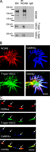
NCAM associates with T- and L-type VDCC. (A) NCAM immunoprecipitates (IP) from NCAM+/+ brain lysates were analyzed by Western blotting with antibodies against NCAM and T- and L-type VDCC. Mock immunoprecipitation with nonspecific IgG served as control. Brain homogenate (BH) is shown for comparison. T- and L-type VDCC coimmunoprecipitate with NCAM. (B) A growth cone of a cultured hippocampal neuron colabeled with antibodies against NCAM, CaMKIIα, and T-type VDCC is shown. Note the colocalization of these three proteins in filopodia. (C) NCAM was clustered at the surface of cultured hippocampal neurons with NCAM monoclonal antibodies (H28live), and neurons were colabeled with antibodies against CaMKIIα and T-type VDCC. A representative neurite is shown. Note that clusters of NCAM overlap with accumulations of T-type VDCC and CaMKIIα (arrows). Bars, 5 μm.
Figure 8.
Figure 8.
NCAM-induced CaMKIIα activation depends on Ca2+ influx via T- and L-type VDCC. (A) Cultured hippocampal neurons were incubated with pimozide and nifedipine, inhibitors of T-and L-type VDCC, respectively. Neurons were then stimulated with rat NCAM monoclonal antibodies (H28live) or nonspecific rat IgG, then fixed and colabeled with antibodies against activated Thr286-phosphorylated CaMKIIα. Representative images are shown. Note that clustering of NCAM without inhibitors induces an increase in active CaMKIIα levels along neurites and that active CaMKIIα accumulates in NCAM clusters. Pimozide and nifedipine inhibit CaMKIIα activation. Graph shows mean levels ± SEM in arbitrary units (AU) of active CaMKIIα along neurites. n > 45 neurites from 30 neurons from 3 coverslips analyzed in each group. Bar, 20 μm. (B) Cultured cortical neurons were treated with nonspecific rabbit IgG or NCAM polyclonal antibodies in the absence or presence of pimozide and nifedipine. Lysates of these cells and fyn immunoprecipitates from the lysates were then probed with the indicated antibodies by Western blotting. Note that pimozide and nifedipine inhibited fyn and CaMKIIα activation in response to NCAM antibodies. Graphs show quantitation of the blots (mean ± SEM), with the levels in cells treated with nonspecific IgG without inhibitors set to 100%. *, P < 0.05 (paired t test).
Figure 9.
Figure 9.
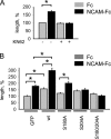
NCAM-induced phosphorylation of RPTPα on Ser180 and Ser204 is required for NCAM-mediated neurite outgrowth. (A) Cultured hippocampal neurons were treated with Fc or NCAM-Fc in the presence or absence of KN62. Graph shows mean lengths of neurites ± SEM normalized to the mean neurite length in neurons treated with Fc in the absence of KN62 (n > 200). Note that KN62 inhibits NCAM-dependent neurite outgrowth. (B) Cultured hippocampal neurons were transfected with GFP alone or cotransfected with GFP and RPTPαWT or RPTPα mutants and treated with Fc or NCAM-Fc. Graph shows mean lengths of neurites ± SEM normalized to the mean neurite length in Fc-treated GFP-transfected neurons (n > 150). Note that RPTPαWT promotes neurite outgrowth and potentiates the response to NCAM-Fc. Mutation in either Ser180 or Ser204 inhibits the outgrowth-promoting effect of RPTPα, whereas the RPTPαS180/204A mutant functions as a dominant-negative construct that fully blocks NCAM-dependent neurite outgrowth. *, P < 0.05 (t test).
Figure 10.
Figure 10.
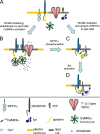
A schematic model of molecular interactions induced by NCAM140 clustering and resulting in fyn activation. (A) Nonclustered NCAM140 resides outside of lipid rafts and does not associate with RPTPα, CaMKIIα (shown as a holoenzyme), and fyn, which are present in inactive forms in the plasma membrane outside of lipid rafts, in the cytosol and in lipid rafts, respectively. (B) NCAM140 clustering induces palmitoylation of the intracellular domain of NCAM140 that promotes NCAM140 redistribution to lipid rafts, where it binds to prion protein (PrP). In parallel, NCAM140 clustering induces FGFR activation, promoting arachidonic acid production (not depicted), which results in arachidonic acid–dependent Ca2+ influx (Williams et al., 1994) via T- and L-type VDCC. An increase in Ca2+ concentration promotes binding of Ca2+/CaM to CaMKIIα, releasing the catalytic domain of CaMKIIα from inhibition by autoregulatory sequences proximal to the CaM binding site (not depicted). By associating with T- and L-type VDCC, NCAM anchors CaMKIIα, which is bound to NCAM140 via spectrin, near the Ca2+ influx sites. NCAM-induced aggregation of CaMKIIα in lipid rafts promotes transautophosphorylation of the CaMKIIα holoenzymes at Thr286, resulting in the constitutive activation of CaMKIIα. (C) In parallel to CaMKIIα activation, NCAM140 promotes redistribution of RPTPα to lipid rafts. In lipid rafts, activated CaMKIIα (only one CaMKIIα molecule is shown for simplicity) phosphorylates RPTPα at Ser180 and Ser204, which changes the conformation of RPTPα, resulting in enzyme activation. (D) Activated RPTPα binds and dephosphorylates fyn at Tyr531, activating the enzyme. Downstream targets of active fyn include the Ras–MAP (MAP) kinase pathway, the sustained activity of which is required for neuronal differentiation (not depicted).

Similar articles

Cited by

References

    1. Beggs, H.E., P. Soriano, and P.F. Maness. 1994. NCAM-dependent neurite outgrowth is inhibited in neurons from Fyn-minus mice. J. Cell Biol. 127:825–833. - PMC - PubMed
    1. Beggs, H.E., S.C. Baragona, J.J. Hemperly, and P.F. Maness. 1997. NCAM140 interacts with the focal adhesion kinase p125(fak) and the SRC-related tyrosine kinase p59(fyn). J. Biol. Chem. 272:8310–8319. - PubMed
    1. Bhandari, V., K.L. Lim, and C.J. Pallen. 1998. Physical and functional interactions between receptor-like protein-tyrosine phosphatase alpha and p59fyn. J. Biol. Chem. 273:8691–8698. - PubMed
    1. Blanchetot, C., and J. den Hertog. 2000. Multiple interactions between receptor protein-tyrosine phosphatase (RPTP) alpha and membrane-distal protein-tyrosine phosphatase domains of various RPTPs. J. Biol. Chem. 275:12446–12452. - PubMed
    1. Bodrikov, V., I. Leshchyns'ka, V. Sytnyk, J. Overvoorde, J. den Hertog, and M. Schachner. 2005. RPTPα is essential for NCAM-mediated p59fyn activation and neurite elongation. J. Cell Biol. 168:127–139. - PMC - PubMed

Publication types

MeSH terms

Substances