. 2008 Sep 22;182(6):1231-44.
doi: 10.1083/jcb.200801121.

The WAVE2 complex regulates T cell receptor signaling to integrins via Abl- and CrkL-C3G-mediated activation of Rap1

Affiliations

The WAVE2 complex regulates T cell receptor signaling to integrins via Abl- and CrkL-C3G-mediated activation of Rap1

Jeffrey C Nolz et al. J Cell Biol. .

Abstract

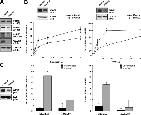
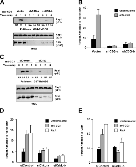
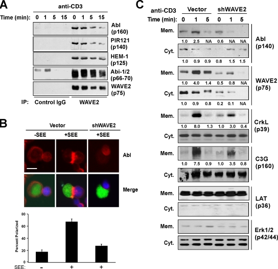
WAVE2 regulates T cell receptor (TCR)-stimulated actin cytoskeletal dynamics leading to both integrin clustering and affinity maturation. Although WAVE2 mediates integrin affinity maturation by recruiting vinculin and talin to the immunological synapse in an Arp2/3-dependent manner, the mechanism by which it regulates integrin clustering is unclear. We show that the Abl tyrosine kinase associates with the WAVE2 complex and TCR ligation induces WAVE2-dependent membrane recruitment of Abl. Furthermore, we show that WAVE2 regulates TCR-mediated activation of the integrin regulatory guanosine triphosphatase Rap1 via the recruitment and activation of the CrkL-C3G exchange complex. Moreover, we demonstrate that although Abl does not regulate the recruitment of CrkL-C3G into the membrane, it does affect the tyrosine phosphorylation of C3G, which is required for its guanine nucleotide exchange factor activity toward Rap1. This signaling node regulates not only TCR-stimulated integrin clustering but also affinity maturation. These findings identify a previously unknown mechanism by which the WAVE2 complex regulates TCR signaling to Rap1 and integrin activation.


Figures

Figure 1.
Figure 1.
WAVE2 is required for TCR-mediated adhesion to both fibronectin and ICAM-1 in primary human CD4+ T cells. (A) Purified human CD4+ T cells were transfected with either control siRNA (siControl) or siRNA against WAVE2 (siWAVE2). Whole cell extracts were then analyzed by immunoblot for expression of WAVE2, Abi-1/2, HEM-1, PIR121, and ZAP-70. (B and C) Cells were transfected as in A, stained with calcein AM, and added to a 96-well plate coated with increasing concentrations of fibronectin or ICAM-1-Fc (B) or 0.3 μg of each (C). Cells were then left unstimulated (C) or stimulated with anti-CD3 (B and C), and adhesion was determined as described in Materials and methods. Error bars represent SD.
Figure 2.
Figure 2.
WAVE2 is required for TCR-mediated Rap1 activation. (A) Jurkat T cells were transfected with either control vector or WAVE2 suppression vector and allowed to recover for 72 h. Cells were then stimulated with anti-CD3 for the indicated time, lysed, and GTP-bound Rap1 was detected using GST-RalGDS GBD and immunoblot. (B and C) Same as A except purified human CD4+ or CD8+ T cells were transfected with either siControl or siWAVE2 and stimulated with anti-CD3 for the indicated times. Black lines indicate that intervening lanes have been spliced out. (D and E) Jurkat T cells were transfected with the indicated vectors and stimulated with either anti-CD3 or 50 ng/ml PMA for 2 min. Subsequently, cytosolic and membrane fractions were prepared and probed as indicated. (F) Purified human CD4+ T cells were transfected as in B, stimulated with anti-CD3 over the indicated timecourse, and cell lysates were immunoblotted with a phospho-specific antibody against PAK (which cross-reacts with Mst1), total Mst1, and WAVE2. Numbers below top blots are arbitrary units based on densitometric analysis of the immunoblots.
Figure 3.
Figure 3.
C3G and CrkL are required for TCR-mediated activation of Rap1 and adhesion. (A) Jurkat T cells were transfected with the indicated suppression vectors, stimulated with anti-CD3, and Rap1 activation was determined. (B) Cells were transfected as in A, stained with calcein AM, and adhesion to 0.3 μg/well fibronectin was determined. (C) Purified human CD4+ T cells were transfected with the indicated siRNAs and Rap1 activation after anti-CD3 cross-linking was determined as described in Materials and methods. Numbers below top blots are arbitrary units based on densitometric analysis of the immunoblots. (D and E) Purified human CD4+ T cells were transfected with the indicated siRNAs and analyzed for the ability to adhere to 0.1 μg/well fibronectin or 0.3 μg/ml ICAM-1-Fc in response to either anti-CD3 or 50 ng/ml PMA. Error bars represent SD.
Figure 4.
Figure 4.
CrkL and C3G regulate both integrin affinity and avidity. (A) Jurkat T cells were transfected with GFP-expressing control or suppression vectors targeting either CrkL or C3G. T cells (green) were allowed to form conjugates with superantigen (SEE)-pulsed Raji B cells (blue) and stained for β1 integrin (red) to determine localization of integrins to the IS. Data shown is the mean of three independent experiments. Bar, 5 μm. (B) Cells were transfected as in A and analyzed for their ability to bind soluble VCAM-1–Fc in response to either anti-CD3 or PMA stimulation. Stimulation with Mn2+ serves as a positive control for VCAM-1–Fc binding. Histograms represent VCAM-1–Fc binding in cell populations gated on different levels of GFP expression (negative, low, and high).
Figure 5.
Figure 5.
WAVE2 is required for phosphorylation of CrkL and C3G. (A) Jurkat T cells were stimulated with OKT3-bound beads, and the localization of WAVE2 (green) or CrkL (green) and F-actin (red) was analyzed by confocal microscopy. Representative images are shown. Bar, 5 μm. (B) Jurkat T cells were transfected with the indicated vectors and stimulated with anti-CD3. Whole cell extracts were prepared and immunoblotted for pCrkL (Y207), total CrkL, and WAVE2. (C) Jurkat T cells were transfected with the indicated suppression/reexpression vectors, stimulated with anti-CD3, and whole cell extracts were immunoblotted as in B. (D) Jurkat T cells were transfected with the indicated expression vectors, stimulated with anti-CD3, and lysates immunoblotted with the indicated antibodies. (E) Jurkat T cells were transfected with control vector, CrkL-suppression vector, or suppression/reexpression vectors, which reexpressed either wild-type CrkL or point mutants disrupting the function of the SH2, SH3-N, or SH3-C domains. Rap1 activation was assessed as previously described. (F) Cells were transfected and stimulated as in B, and C3G was purified using a GST-CrkL SH3-N fusion protein and subsequently blotted using the indicated antibodies. (G) Purified human CD4+ T cells were transfected with the indicated siRNAs, stimulated with anti-CD3, and Rap1 activation and phosphorylation of CrkL was analyzed by immunoblot. Numbers below top blots are arbitrary units based on densitometric analysis of the immunoblots. Black lines indicate that intervening lanes have been spliced out.
Figure 6.
Figure 6.
The Abl tyrosine kinase interacts with and is localized to the membrane by the WAVE2 complex. (A) Purified human T cells were stimulated with anti-CD3. WAVE2 was immunoprecipitated and bound proteins were immunoblotted as indicated. (B) Control or WAVE2-suppressed Jurkat T cells (green) were allowed to form conjugates with antigen-pulsed Nalm 6 B cells (blue) and then stained for Abl (red). The percentage of T–B cell conjugates showing Abl accumulation at the IS were quantified as described in the Materials and methods. Error bars represent SD. Bar, 5 μm. (C) Jurkat T cells were transfected with either control or WAVE2 suppression vector and stimulated with anti-CD3. Subsequently, cytosolic and membrane fractions were collected as described in Materials and methods and immunoblotted with the indicated antibodies. Numbers below blots are arbitrary units based on densitometric analysis of the immunoblots.
Figure 7.
Figure 7.
Abl regulates TCR-mediated activation of Rap1 and integrin affinity maturation. (A) Purified human CD4+ T cells were transfected with the indicated siRNAs, and Rap1 activation was assessed after anti-CD3 cross-linking. Numbers below top blot are arbitrary units based on densitometric analysis of the immunoblots. (B) Same as A, except whole cell extracts were collected and analyzed by immunoblot for pMst1, Mst1, and Abl. (C) Jurkat T cells were transfected with suppression vectors against Abl (Abl-1 and Abl-3), stimulated with anti-CD3, and cell extracts were analyzed for pPLCγ1 (Y783), PLCγ1, pCrkL (Y207), CrkL, and Abl. (D and E) Jurkat T cells (D) or primary human CD4+ T cells (E) were suppressed for Abl and basal, and anti-CD3 and PMA-stimulated adhesion to fibronectin was analyzed as described in Fig. 1. Error bars represent SD. (F) Jurkat T cells were transfected with the indicated GFP-expressing suppression vector cells and analyzed for their ability to bind soluble VCAM-1-Fc as in Fig. 4 B.
Figure 8.
Figure 8.
Affect of Abl suppression on CrkL–C3G membrane localization and activation. (A) Jurkat T cells transfected with control, WAVE2, or Abl EGFP suppression vectors were stimulated with IgG (column 1) or OKT3-coated beads (columns 2–4) and imaged for CrkL (aqua) and F-actin (red) recruitment. The percentage of EGFP+ T cell–bead conjugates showing localization of CrkL is indicated and was performed as described in Materials and methods. Representative images are shown. Bar, 5 μm. (B) Jurkat T cells were transfected with the indicated suppression vectors, stimulated by anti-CD3 cross-linking and cytosolic/membrane fractions, were prepared and immunoblotted with the indicated antibodies. Numbers below blots are arbitrary units based on densitometric analysis of the immunoblots. (C) Purified Abl and Fyn were incubated with the indicated GST fusion proteins in kinase buffer, and tyrosine phosphorylation was detected by anti-Tyr immunoblotting. Input levels of GST were detected by anti-GST immunoblotting. Numbers on the left are arbitrary units based on densitometric analysis of the immunoblots. (D) Jurkat T cells were transfected with the indicated suppression vectors, stimulated by anti-CD3 cross-linking, and C3G was precipitated as described in Fig. 5 E. Proteins were detected by immunoblotting with the indicated antibodies. Numbers below blots are arbitrary units based on densitometric analysis of the immunoblots.
Figure 9.
Figure 9.
Suppression of WAVE2, Abl, CrkL, and C3G affects IL-2 production in T cells. Primary human CD4+ T cells were transfected with the indicated siRNA duplexes. After a 72-h incubation, cells were left unstimulated or stimulated with Nalm 6 B cells loaded with or without superantigen cocktail. 15 h after incubation, the amount or IL-2 in the supernatant was determined by ELISA as described in the Materials and methods. Error bars represent SD.

Similar articles

Cited by

References

    1. Anton van der Merwe, P., S.J. Davis, A.S. Shaw, and M.L. Dustin. 2000. Cytoskeletal polarization and redistribution of cell-surface molecules during T cell antigen recognition. Semin. Immunol. 12:5–21. - PubMed
    1. Arai, A., Y. Nosaka, H. Kohsaka, N. Miyasaka, and O. Miura. 1999. CrkL activates integrin-mediated hematopoietic cell adhesion through the guanine nucleotide exchange factor C3G. Blood. 93:3713–3722. - PubMed
    1. Billadeau, D.D., J.C. Nolz, and T.S. Gomez. 2007. Regulation of T-cell activation by the cytoskeleton. Nat. Rev. Immunol. 7:131–143. - PubMed
    1. Bos, J.L. 2005. Linking Rap to cell adhesion. Curr. Opin. Cell Biol. 17:123–128. - PubMed
    1. Cannon, J.L., C.M. Labno, G. Bosco, A. Seth, M.H. McGavin, K.A. Siminovitch, M.K. Rosen, and J.K. Burkhardt. 2001. Wasp recruitment to the T cell:APC contact site occurs independently of Cdc42 activation. Immunity. 15:249–259. - PubMed

Publication types

MeSH terms