. 2008 Oct 7;105(40):15399-404.
doi: 10.1073/pnas.0807301105. Epub 2008 Sep 22.

Loss of p120 catenin and links to mitotic alterations, inflammation, and skin cancer

Affiliations

Loss of p120 catenin and links to mitotic alterations, inflammation, and skin cancer

Mirna Perez-Moreno et al. Proc Natl Acad Sci U S A. .

Abstract

Tumor formation involves epigenetic modifications and microenvironmental changes as well as cumulative genetic alterations encompassing somatic mutations, loss of heterozygosity, and aneuploidy. Here, we show that conditional targeting of p120 catenin in mice leads to progressive development of skin neoplasias associated with intrinsic NF-kappaB activation. We find that, similarly, squamous cell carcinomas in humans display altered p120 and activated NF-kappaB. We show that epidermal hyperproliferation arising from p120 loss can be abrogated by IkappaB kinase 2 inhibitors. Although this underscores the importance of this pathway, the role of NF-kappaB in hyperproliferation appears rooted in its impact on epidermal microenvironment because as p120-null keratinocytes display a growth-arrested phenotype in culture. We trace this to a mitotic defect, resulting in unstable, binucleated cells in vitro and in vivo. We show that the abnormal mitoses can be ameliorated by inhibiting RhoA, the activity of which is abnormally high. Conversely, we can elicit such mitotic defects in control keratinocytes by elevating RhoA activity. The ability of p120 deficiency to elicit mitotic alterations and chronic inflammatory responses, that together may facilitate the development of genetic instability in vivo, provides insights into why it figures so prominently in skin cancer progression.


Conflict of interest statement

The authors declare no conflict of interest.

Figures

Fig. 1.
Fig. 1.
Absence of p120 in skin promotes tumor formation. WT and p120 cKO newborn back skins were engrafted onto nude mice. (A) Phenotypic appearance of skin grafts. Note that 50-d cKO grafts display hyperkeratosis and a paucity of hair. At later stages, grafts appear as a group of papules or nodules with various degrees of hyperkeratosis and ulceration. (B and C) Histological analyses show that 50-d cKO grafts exhibit well circumscribed, keratin-filled, epidermal invaginations that extend into the dermis. Signs of hyperkeratosis and acanthosis with atypical cells are evident. By 70 d, cKO grafts display some signs of dysplasia and early neoplasia. Immunohistochemistry reveals expansion of K5 expression. (D–G) Immunofluorescence was conducted on frozen sections of 50-d (D and E) and 70-d (F and G) grafts with the indicated Abs. Notes on cKO grafts: AJ proteins no longer localize to cell borders, cKO epidermis is hyperproliferative, the basement membrane is largely intact, and the TGFβ effector nuclear pSmad2 is prominent. Abbreviations: Epi, epidermis; der, dermis; hf, hair follicles; E-cad, E-cadherin; β4, β4 integrin; Ker, keratin; Lam, Laminin; bv, blood vessel.
Fig. 2.
Fig. 2.
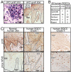
p120 loss, NF-κB activation, and relationship with human skin cancer. (A) Histologic and immunohistochemistry analyses of skin grafts reveal an association of p120 loss with chronic inflammatory infiltrates (arrowheads) and NF-κB phosphorylation/activation. (B and C) Immunohistochemical analyses of p120 and NF-κB in human SCCs of different grades. Table summarizes expression and distribution for 40 human SCCs (the two analyses are presented as independent datasets). Shown are representative examples of the data. Insets denote magnified areas showing well and poorly differentiated regions of a grade I tumor. Note that, in areas where p120 is not detected, NF-κB is nuclear and activated. Abbreviations: Phos, phosphorylated; SQCC, squamous cell carcinoma.
Fig. 3.
Fig. 3.
Inflammatory responses are causative of hyperproliferation features in vivo, whereas in vitro loss of p120 results in hypoproliferation. (A) Histological analyses of skin grafts with and without treatment with an IKK2 inhibitor that specifically prevents NF-κB phosphorylation and activation (n = 5). (B) Blocking NF-κB activation and inflammation rescues hyperproliferation but not cadherin distribution at cell–cell boundaries. (C) Quantification of BrdU incorporation. Error bars indicate SD. (D) TUNEL assay. Note that IKK2 inhibition shows no increase in apoptosis. (E) Ras pull-down assays. Note the small decrease on the levels of active Ras in p120 cKO neonatal epidermis. (F–I). In vitro studies. (F) Proliferation curves of p120-null mKer with and without IKK2 inhibitor treatment. Note that p120 loss, but not IKK2 inhibition, affects mKer proliferation in vitro. (G) Colony formation efficiency after 10 d of plating. (H) TUNEL assay with and without IKK2 inhibitor. (I) Adhesion assays on different extracellular matrices.
Fig. 4.
Fig. 4.
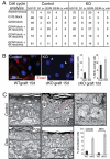
Loss of p120 leads to the generation of binucleated cells in vivo and in vitro. (A) FACS analysis of DNA content profile of asynchronously growing mKers and of G1/S and G2/M synchronized cultures (see Methods for details). (B Left) Examples of binucleate cells in p120-null mKers (arrowheads; red, E-cad; blue, DAPI). (Right) Quantification of binucleate cells and S-phase (i.e., BrdU-labeled) cells in control and p120-null cultures. (C) Morphological analyses of skin grafts processed for Epon embedding and sectioning. Note that binucleated epidermal cells (arrowheads) appear in p120 cKO skin even when inflammation was blocked with dexamethasone. Inset shows magnified view of boxed area. Abbreviations: cv, coefficient of variation; Dex, dexamethasone.
Fig. 5.
Fig. 5.
p120-null cells exhibit abnormal mitosis in a RhoA GTPase-dependent fashion. (A) Quantification of the extended length of M-phase in KO mKers. Mitoses were monitored by bright-field videoimaging. Shown are control (n = 60) and p120-null mKers (n = 58). Mean times of total mitoses are shown. (B) Immunofluorescence microscopy of mKers labeled with Abs is indicated (color coding according to the secondary). Note the presence of abnormal spindles and centrosomes in KO cells. Black-and-white images depict a representative example of lagging chromosomes and chromosomal bridges in p120-null cells (arrows). (C) RhoA activity is increased in newborn p120 cKO epidermis. (D) Percentage of cells with normal and abnormal mitosis (i.e., binucleated cells, furrow regression, and mitotic catastrophe) and dependency on RhoA activity. Abbreviations: H2B, histone 2B; DA, dominant active; DN, dominant negative.

Comment in

Similar articles

Cited by

References

    1. Reynolds AB. p120-catenin: Past and present. Biochim Biophys Acta. 2007;1773:2–7. - PMC - PubMed
    1. Ireton RC, et al. A novel role for p120 catenin in E-cadherin function. J Cell Biol. 2002;159:465–476. - PMC - PubMed
    1. Davis MA, Ireton RC, Reynolds AB. A core function for p120-catenin in cadherin turnover. J Cell Biol. 2003;163:525–534. - PMC - PubMed
    1. Birchmeier W, Behrens J. Cadherin expression in carcinomas: Role in the formation of cell junctions and the prevention of invasiveness. Biochim Biophys Acta. 1994;1198:11–26. - PubMed
    1. Reynolds AB, Roczniak-Ferguson A. Emerging roles for p120-catenin in cell adhesion and cancer. Oncogene. 2004;23:7947–7956. - PubMed

Publication types

MeSH terms