. 2008 Sep 30;105(39):15016-21.
doi: 10.1073/pnas.0801497105. Epub 2008 Sep 22.

Selective expansion of a subset of exhausted CD8 T cells by alphaPD-L1 blockade

Affiliations

Selective expansion of a subset of exhausted CD8 T cells by alphaPD-L1 blockade

Shawn D Blackburn et al. Proc Natl Acad Sci U S A. .

Abstract

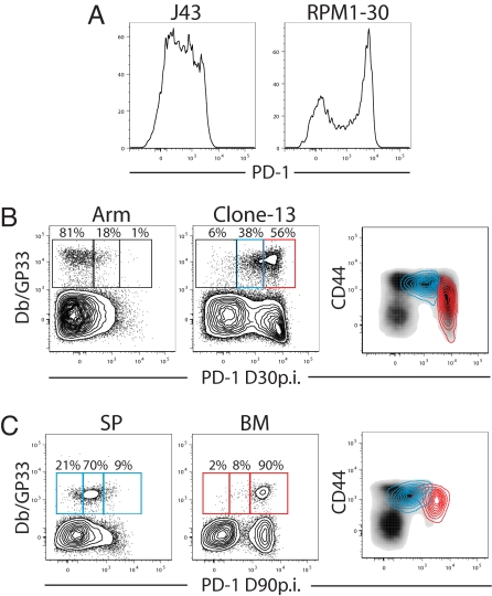
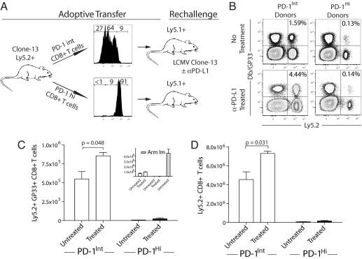
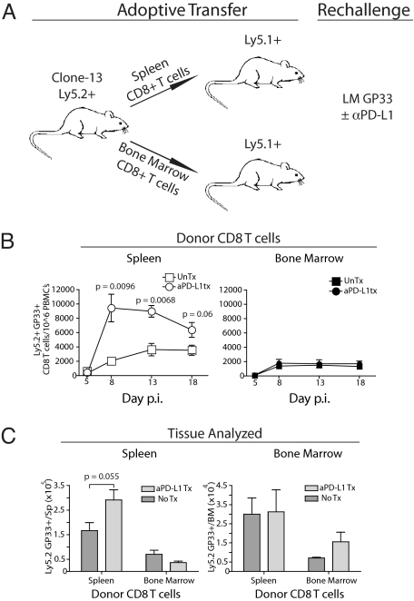
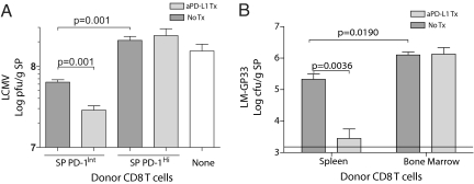
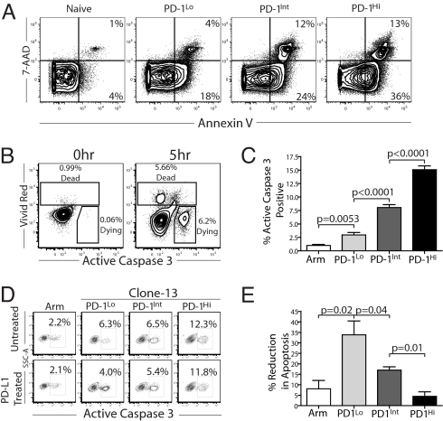
Programmed death-1 (PD-1) regulates T cell exhaustion during chronic infections. Blocking the PD-1:PD-ligand (PD-L) pathway reinvigorates exhausted CD8 T cells. Exactly how blocking PD-1:PD-L interactions improves T cell immunity, however, remains unclear. PD-1:PD-L blockade could reprogram all exhausted T cells to become antiviral effectors. Alternatively, this blockade might selectively expand a subset of exhausted T cells. We have identified two subpopulations of exhausted CD8 T cells during chronic viral infection in mice. One subset of exhausted CD8 T cells is rescued by alphaPD-L1 blockade, whereas the other subset appears more terminally differentiated and responds poorly to PD-1:PD-L blockade. Blocking PD-1:PD-L interactions reduces spontaneous apoptosis and enhances expansion and protective immunity of the rescuable subset, but not the more terminally differentiated subset of exhausted CD8 T cells. These results have implications for predicting clinical responses to PD-1-based therapeutic interventions and for understanding T cell dynamics during persisting infections.


Conflict of interest statement

The authors declare no conflict of interest.

Figures

Fig. 1.
Fig. 1.
Identification of subsets of exhausted CD8 T cells. (A) CD8+ CD44+ T cells from Arm vs. clone 13-infected mice (day 30 after infection) were stained with either the J43 clone or the RPM1–30 clone of αPD-1. (B) Splenocytes from either LCMV Arm or clone 13-infected mice (day 30 after infection) were stained with αPD-1 clone RPM1–30 and gated on CD8+ T cells. PD-1Hi (red) and PD-1Int (blue) DbGP33+ CD8+ T cells were overlaid on total CD8 T cells (black; Right) from the same mice to assess overlap of these populations and to illustrate differences in CD44 expression. (C) Lymphocytes from SP or BM of clone 13-infected mice (day 90 after infection) were stained with αPD-1 clone RPM1–30. Plots are gated on CD8+ T cells. DbGP33+ CD8+ T cells from the SP or BM. DbGP33+ CD8+ T cells from SP (blue) or BM (red) were overlaid total CD8+ splenocytes (black; Right).
Fig. 2.
Fig. 2.
In vivo PD-L1 blockade enhanced expansion of splenic PD-1Int, but not the PD-1Hi subset of exhausted CD8 T cells. (A) PD-1Hi and PD-1Int splenocytes from clone 13-infected (day 30 after infection) Ly5.2+ mice were column-purified. Each fraction yielded >90% purity for PD-1Hi and PD-1Int. Note that the PD-1Int fraction contained a small percentage of PD-1Lo cells, but these cells expressed higher PD-1 levels compared with memory CD8 T cells. DbGP33+ CD8+ T cells (6 × 10⁴) were adoptively transferred to Ly5.1 naïve recipient mice followed by rechallenge with LCMV clone 13 with or without αPD-L1 antibody treatment (200 μg i.p. on days 0, 3, and 6 after infection). As a control, LCMV-specific memory CD8 T cells from Ly5.2+ LCMV Arm immune mice were transferred to Ly5.1+ naïve recipients and rechallenged. (B–D) On day 7.5 after rechallenge, recipient mice were killed, and total donor Ly5.2+ or DbGP33+ Ly5.2+ CD8+ T cells were identified by flow cytometry (n = 3–5 mice per group, data are representative of three similar experiments).
Fig. 3.
Fig. 3.
In vivo PD-L1 blockade enhanced expansion of tissue-defined PD-1Int (SP-derived), but not the PD-1Hi (BM-derived) subset of exhausted CD8 T cells. (A) PD-1Hi CD8+ T cells were derived from the BM and PD-1Int CD8+ T cells from the SP of clone 13-infected (day 90 after infection) Ly5.2+ mice. CD8 T cells from BM and SP were column-purified, CD8+ T cell populations were normalized to contain 6 × 10⁴ DbGP33+ CD8+T cells, and cells were adoptively transferred to Ly5.1 naïve recipient mice followed by rechallenge with LMGP33 (3 × 10⁴ cfu) with or without αPD-L1 antibody treatment (200 μg i.p. on days 0, 3, and 6 after infection). (B) Ly5.2+ DbGP33+ CD8+ T cells were monitored in the blood of recipient mice after LMGP33 rechallenge (n = 3–5 mice per group, data are representative of five experiments). (C) Total donor DbGP33+ Ly5.2+ CD8+ T cell number was assessed in recipient SP and BM on day 18 after challenge.
Fig. 4.
Fig. 4.
Protective immunity by subsets of exhausted CD8 T cells in the absence or presence PD-L1 blockade. (A) PD-1Hi and PD-1Int splenocytes were purified as in Fig. 2, and 1 × 10⁶ LCMV-specific CD8+ T cells [normalized based on responsiveness via ICS to a pool of 20 known LCMV CD8 T cell epitopes described by Kotturi et al. (24)] were transferred to Ly5.1 naïve recipient mice followed by rechallenge with LCMV clone 13 (2 × 10⁶pfu i.v.) with or without αPD-L1 antibody treatment as in Fig. 2. As a control, Ly5.1+ mice that received no donors cells were also infected. Mice were killed on day 3 after infection. and LCMV quantified in the SP. (B) Adoptive transfers were performed as described in Fig. 3. Recipients were then rechallenged with LMGP33 (1.5 × 10⁴ cfu i.v.) with or without αPD-L1 antibody (200 μg i.p. on days 0 and 3 after infection). Mice were killed on day 4 after infection, and LMGP33 was quantified in the SP.
Fig. 5.
Fig. 5.
Differential inhibition of apoptosis of exhausted CD8 T cell subsets by PD-1:PD-L blockade. (A) Splenocytes from clone 13-infected mice (day 30 after infection) were isolated and stained with 7-AAD and annexin V directly ex vivo. (B) ACasp3 was used as an indicator of apoptosis in live cells. Dead cells were excluded by using an amine-reactive dye (vivid red), and ACasp3 was detected by intracellular staining. (C) Splenocytes from clone 13-infected mice were incubated at 37°C for 5 h in medium. ACasp3 was detected in live DbGP33+ CD8 T cells. (D and E) PD-1Hi, PD-1Int, and PD-1Lo exhausted CD8 T cells subsets were sorted by flow cytometry and incubated at 37°C for 5 h with or without αPD-L1 (10 μg/ml), and ACasp3 was detected in the live cell subset by flow cytometry (data are representative of three independent experiments). Note that PD-1 expression did not change in 5 h in vitro (data not shown). Similar results were observed in the presence of added peptide (data not shown).

Similar articles

Cited by

References

    1. Sharpe AH, Wherry EJ, Ahmed R, Freeman GJ. The function of programmed cell death 1 and its ligands in regulating autoimmunity and infection. Nat Immunol. 2007;8:239–245. - PubMed
    1. Barber DL, et al. Restoring function in exhausted CD8 T cells during chronic viral infection. Nature. 2006;439:682–687. - PubMed
    1. Iwai Y, Terawaki S, Ikegawa M, Okazaki T, Honjo T. PD-1 inhibits antiviral immunity at the effector phase in the liver. J Exp Med. 2003;198:39–50. - PMC - PubMed
    1. Isogawa M, Furuichi Y, Chisari FV. Oscillating CD8(+) T cell effector functions after antigen recognition in the liver. Immunity. 2005;23:53–63. - PubMed
    1. Day CL, et al. PD-1 expression on HIV-specific T cells is associated with T cell exhaustion and disease progression. Nature. 2006;443:350–354. - PubMed

Publication types

MeSH terms