

Evaluation of MR markers that predict survival in patients with newly diagnosed GBM prior to adjuvant therapy
- PMID: 18810326
- PMCID: PMC3022437
- DOI: 10.1007/s11060-008-9685-3
Evaluation of MR markers that predict survival in patients with newly diagnosed GBM prior to adjuvant therapy
Abstract
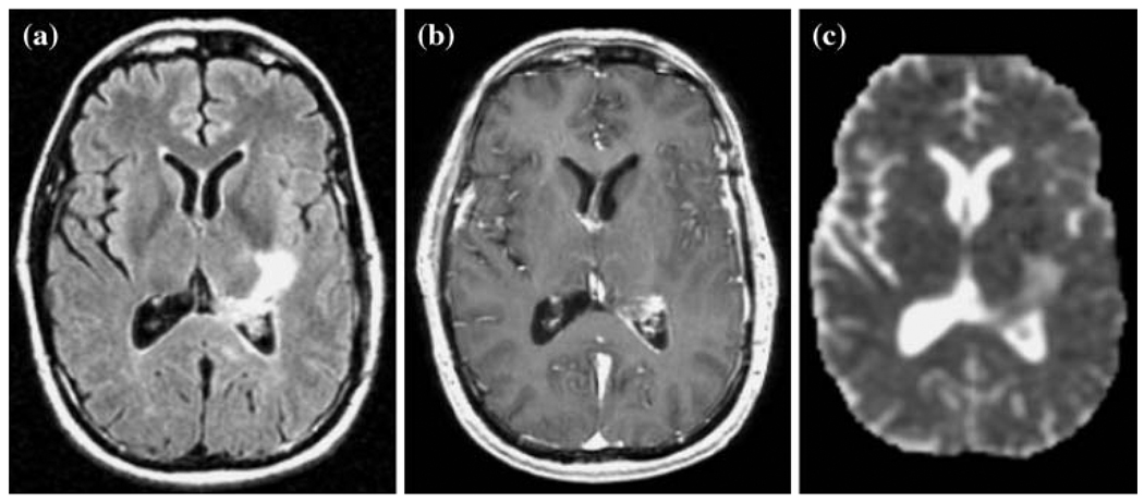
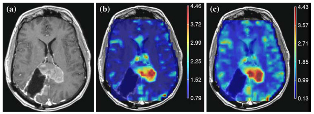
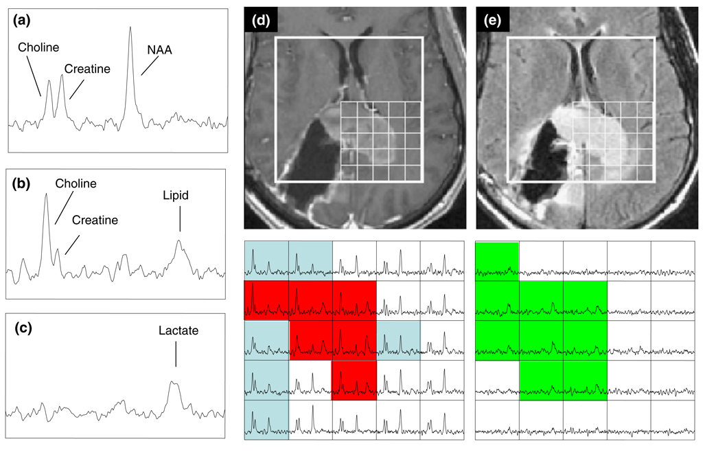
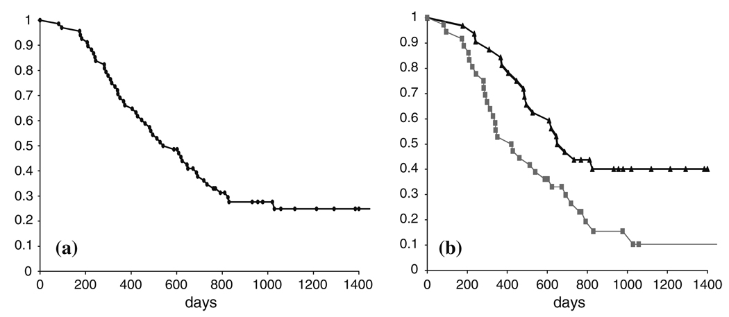
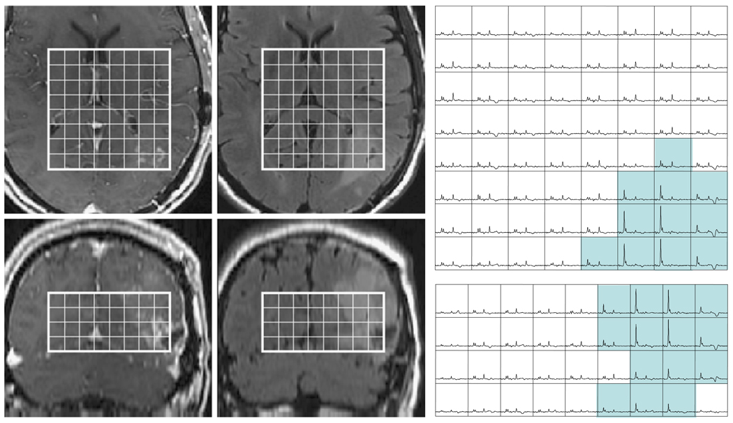
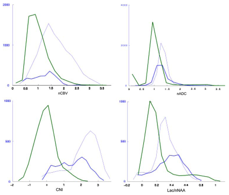
Purpose Glioblastoma Multiforme (GBM) is the most common and lethal primary brain tumor in adults. The goal of this study was to test the predictive value of MR parameters in relation to the survival of patients with newly diagnosed GBM who were scanned prior to receiving adjuvant radiation and chemotherapy. Methods The study population comprised 68 patients who had surgical resection and were to be treated with fractionated external beam radiation therapy and chemotherapy. Imaging scans included anatomical MRI, diffusion and perfusion weighted imaging and (1)H MRSI. The MR data were acquired 3-5 weeks after surgery and approximately 1 week before treatment with radiation therapy. The diffusion, perfusion and spectroscopic parameter values were quantified and subjected to proportional hazards analysis that was adjusted for age and scanner field strength. Results The patients with larger lesion burden based upon volumes of anatomic lesions, volume of CNI2 (number of voxels within the T2 lesion having choline to NAA index >2), volume of CBV3 (number of pixels within the T2 lesion having relative cerebral blood volume >3), and volume of nADC1.5 (number of pixels within the T2 lesion having normalized apparent diffusion coefficient <1.5) had a higher risk for poor outcome. High intensities of combined measures of lactate and lipid in the T2 and CNI2 regions were also associated with poor survival. Conclusions Our study indicated that several pre-treatment anatomic, physiological and metabolic MR parameters are predictive of survival. This information may be important for stratifying patients to specific treatment protocols and for planning focal therapy.
Figures
Similar articles
-
Association of early changes in 1H MRSI parameters with survival for patients with newly diagnosed glioblastoma receiving a multimodality treatment regimen.Neuro Oncol. 2017 Mar 1;19(3):430-439. doi: 10.1093/neuonc/now159. Neuro Oncol. 2017. PMID: 27576874 Free PMC article.
-
Relationship of pre-surgery metabolic and physiological MR imaging parameters to survival for patients with untreated GBM.J Neurooncol. 2009 Feb;91(3):337-51. doi: 10.1007/s11060-008-9719-x. Epub 2008 Nov 15. J Neurooncol. 2009. PMID: 19009235 Free PMC article.
-
Multiparametric MR Imaging of Diffusion and Perfusion in Contrast-enhancing and Nonenhancing Components in Patients with Glioblastoma.Radiology. 2017 Jul;284(1):180-190. doi: 10.1148/radiol.2017160150. Epub 2017 Feb 27. Radiology. 2017. PMID: 28240563
-
Serial analysis of 3D H-1 MRSI for patients with newly diagnosed GBM treated with combination therapy that includes bevacizumab.J Neurooncol. 2016 Oct;130(1):171-179. doi: 10.1007/s11060-016-2229-3. Epub 2016 Aug 17. J Neurooncol. 2016. PMID: 27535746 Free PMC article.
-
The role of radiation therapy in treatment of adults with newly diagnosed glioblastoma multiforme: a systematic review and evidence-based clinical practice guideline update.J Neurooncol. 2020 Nov;150(2):215-267. doi: 10.1007/s11060-020-03612-7. Epub 2020 Nov 19. J Neurooncol. 2020. PMID: 33215344
Cited by
-
Lactylation in Glioblastoma: A Novel Epigenetic Modifier Bridging Epigenetic Plasticity and Metabolic Reprogramming.Int J Mol Sci. 2025 Apr 4;26(7):3368. doi: 10.3390/ijms26073368. Int J Mol Sci. 2025. PMID: 40244246 Free PMC article. Review.
-
MR-Guided Radiotherapy for Brain and Spine Tumors.Front Oncol. 2021 Mar 8;11:626100. doi: 10.3389/fonc.2021.626100. eCollection 2021. Front Oncol. 2021. PMID: 33763361 Free PMC article. Review.
-
Integration method of 3D MR spectroscopy into treatment planning system for glioblastoma IMRT dose painting with integrated simultaneous boost.Radiat Oncol. 2013 Jan 2;8:1. doi: 10.1186/1748-717X-8-1. Radiat Oncol. 2013. PMID: 23280007 Free PMC article.
-
Hypoxia-Driven Immunosuppressive Metabolites in the Tumor Microenvironment: New Approaches for Combinational Immunotherapy.Front Immunol. 2018 Jul 16;9:1591. doi: 10.3389/fimmu.2018.01591. eCollection 2018. Front Immunol. 2018. PMID: 30061885 Free PMC article. Review.
-
Spectroscopic imaging of D-2-hydroxyglutarate and other metabolites in pre-surgical patients with IDH-mutant lower-grade gliomas.J Neurooncol. 2022 Aug;159(1):43-52. doi: 10.1007/s11060-022-04042-3. Epub 2022 Jun 8. J Neurooncol. 2022. PMID: 35672531 Free PMC article.
References
-
- Aboagye EO, Kelson AB, Tracy M, Workman P. Preclinical development and current status of the fluorinated 2-nitroimidazole hypoxia probe N-(2-hydroxy-3,3,3-trifluoropropyl)-2-(2-nitro-1-imidazolyl) acetamide (SR 4554, CRC 94/17): a non-invasive diagnostic probe for the measurement of tumor hypoxia by magnetic resonance spectroscopy and imaging, and by positron emission tomography. Anticancer Drug Des. 1998;13:703–730. - PubMed
-
- Alger JR, Frank JA, Bizzi A, Fulham MJ, DeSouza BX, Duhaney MO, et al. Metabolism of human gliomas: assessment with H-1 MR spectroscopy and F-18 flourodeoxyglucose PET. Radiology. 1990;177:633–641. - PubMed
-
- Basser PJ, Pierpaoli C. Microstructural and physiological features of tissues elucidated by quantitative-diffusion-tensor MRI. J Magn Reson B. 1996;111:209–219. doi: 10.1006/jmrb.1996.0086. - DOI - PubMed
Publication types
MeSH terms
Substances
Grants and funding
LinkOut - more resources
Full Text Sources
Medical
