

Low grade epithelial stromal tumour of the seminal vesicle
- PMID: 18811925
- PMCID: PMC2564931
- DOI: 10.1186/1477-7819-6-101
Low grade epithelial stromal tumour of the seminal vesicle
Abstract
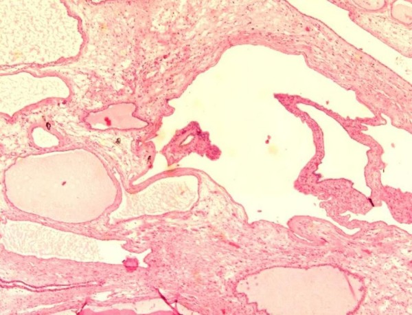
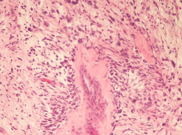
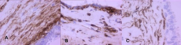
Background: The mixed epithelial stromal tumour is morphologically characterised by a mixture of solid and cystic areas consisting of a biphasic proliferation of glands admixed with solid areas of spindle cells with variable cellularity and growth patterns. In previous reports the seminal vesicle cystoadenoma was either considered a synonym of or misdiagnosed as mixed epithelial stromal tumour. The recent World Health Organisation Classification of Tumours considered the two lesions as two distinct neoplasms. This work is aimed to present the low-grade epithelial stromal tumour case and the review of the literature to the extent of establishing the true frequency of the neoplasm.
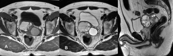
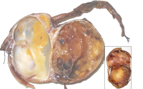
Case presentation: We describe a low-grade epithelial stromal tumour of the seminal vesicle in a 50-year-old man. Computed tomography showed a 9 x 4.5 cm pelvic mass in the side of the seminal vesicle displacing the prostate and the urinary bladder. Magnetic resonance was able to define tissue planes between the lesion and the adjacent structures and provided useful information for an accurate conservative laparotomic surgical approach. The histology revealed biphasic proliferation of benign glands admixed with stromal cellularity, with focal atypia. After 26 months after the excision the patient is still alive with no evidence of disease.
Conclusion: Cystoadenoma and mixed epithelial stromal tumour of seminal vesicle are two distinct pathological entities with different histological features and clinical outcome. Due to the unavailability of accurate prognostic parameters, the prediction of the potential biological evolution of mixed epithelial stromal tumour is still difficult. In our case magnetic resonance imaging was able to avoid an exploratory laparotomy and to establish an accurate conservative surgical treatment of the tumour.
Figures
Similar articles
-
Mixed epithelial and stromal tumor of the seminal vesicles: report of a rare case with diagnostic, therapeutic, and prognostic insights.Diagn Pathol. 2025 Jun 20;20(1):75. doi: 10.1186/s13000-025-01647-w. Diagn Pathol. 2025. PMID: 40542384 Free PMC article.
-
Mixed epithelial-stromal tumor (MEST) of seminal vesicle: a proposal for unified nomenclature.Adv Anat Pathol. 2015 Mar;22(2):113-20. doi: 10.1097/PAP.0000000000000057. Adv Anat Pathol. 2015. PMID: 25664946 Review.
-
Phyllodes tumor of the seminal vesicle: case report and literature review.Pathol Int. 2004 Dec;54(12):924-9. doi: 10.1111/j.1440-1827.2004.01779.x. Pathol Int. 2004. PMID: 15598315
-
Seminal vesicle cystadenoma: a case report and literature review.Urology. 1998 May;51(5):840-5. doi: 10.1016/s0090-4295(97)00711-5. Urology. 1998. PMID: 9610606 Review.
-
Large phyllodes tumour of the seminal vesicle: case report and literature review.J Int Med Res. 2010 Sep-Oct;38(5):1861-7. doi: 10.1177/147323001003800534. J Int Med Res. 2010. PMID: 21309503
Cited by
-
Importance of an Early Diagnosis in Primary Adenocarcinoma of the Seminal Vesicle.Rare Tumors. 2016 Apr 6;8(1):6187. doi: 10.4081/rt.2016.6187. eCollection 2016 Mar 21. Rare Tumors. 2016. PMID: 27134716 Free PMC article.
-
Mixed epithelial and stromal tumor of the seminal vesicles: report of a rare case with diagnostic, therapeutic, and prognostic insights.Diagn Pathol. 2025 Jun 20;20(1):75. doi: 10.1186/s13000-025-01647-w. Diagn Pathol. 2025. PMID: 40542384 Free PMC article.
-
Seminal vesicle schwannoma presenting with left hydroureteronephrosis.Urol Ann. 2014 Oct;6(4):363-5. doi: 10.4103/0974-7796.141007. Urol Ann. 2014. PMID: 25371618 Free PMC article.
-
Incidental early mixed epithelial and stromal tumor of the efferent testicular-ductular system of the genitourinary tract: A small case series with literature review.Pathol Res Pract. 2025 Apr;268:155797. doi: 10.1016/j.prp.2024.155797. Epub 2025 Feb 13. Pathol Res Pract. 2025. PMID: 39983275 Review.
-
Massive mixed epithelial-stromal tumour of seminal vesicle requiring challenging abdominoperineal resection: a case report and review of literature.J Surg Case Rep. 2023 Aug 29;2023(8):rjad490. doi: 10.1093/jscr/rjad490. eCollection 2023 Aug. J Surg Case Rep. 2023. PMID: 37662445 Free PMC article.
References
-
- Eble JN, Sauter G, Epstein JI, Sesterhenn IA, (eds) Pathology and genetics of tumours of the urinary system and male genital organs World Health Organisation Classification of Tumours. IARC Press, Lyon; 2004. pp. 214–215.
-
- Soule EH, Dockerty MB. Cystadenoma of the seminal vesicle, a pathologic curiosity. Report of a case and review of the literature concerning benign tumours of the seminal vesicle. Staff Meet Mayo Clin. 1951;26:406–16. - PubMed
-
- Damjanov I, Apic R. Cystadenoma of seminal vesicles. J Urol. 1974;111:808–809. - PubMed
-
- Lundhus E, Bundgaard N, Sorensen FB. Cystadenoma of the seminal vesicle. Scand J Urol Nephrol. 1984;18:341–342. - PubMed
Publication types
MeSH terms
LinkOut - more resources
Full Text Sources
Molecular Biology Databases
