. 2008 Sep 23:9:25.
doi: 10.1186/1471-2091-9-25.

The Serine/threonine kinase Stk33 exhibits autophosphorylation and phosphorylates the intermediate filament protein Vimentin

Affiliations

The Serine/threonine kinase Stk33 exhibits autophosphorylation and phosphorylates the intermediate filament protein Vimentin

Bastienne Brauksiepe et al. BMC Biochem. .

Abstract

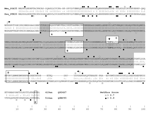
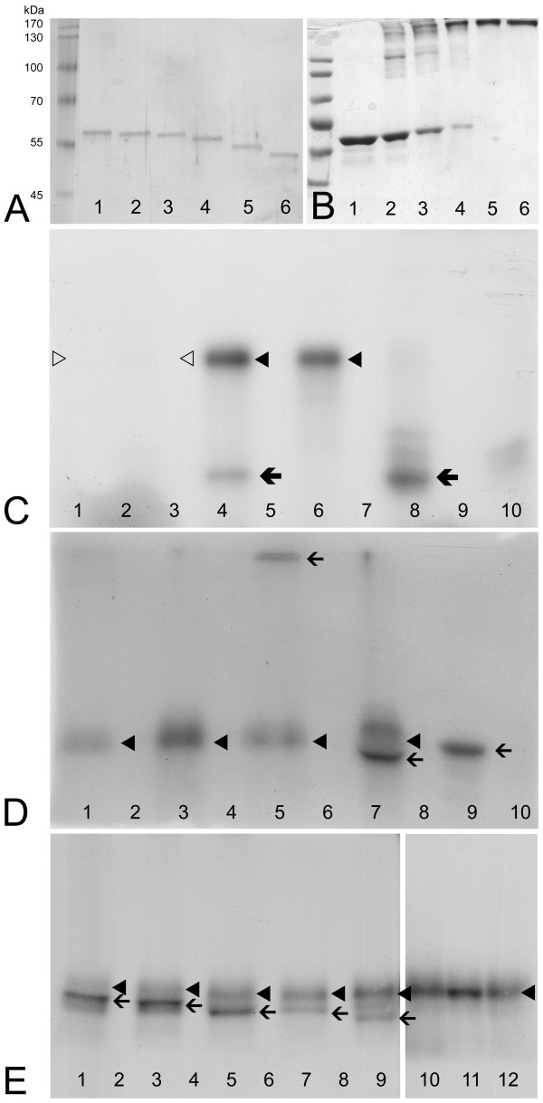
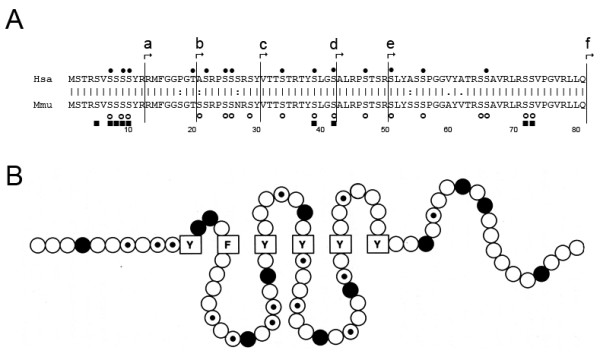
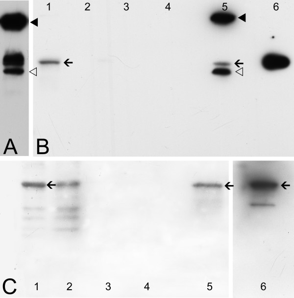
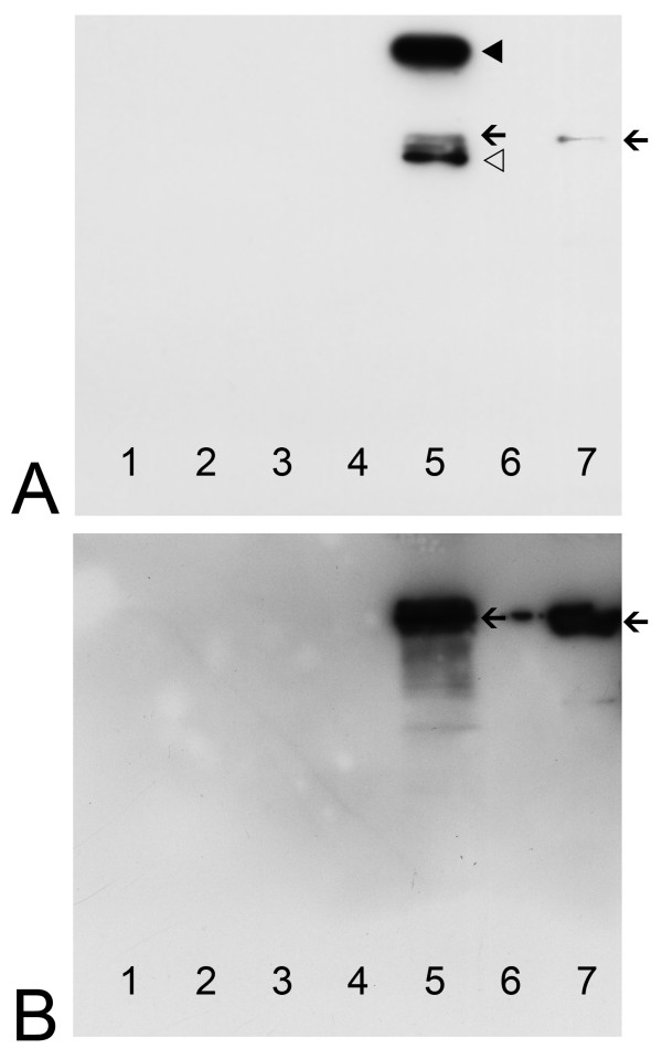
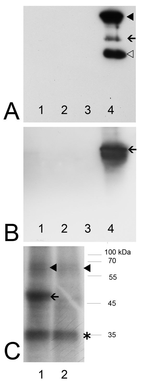
Background: Colocalization of Stk33 with vimentin by double immunofluorescence in certain cells indicated that vimentin might be a target for phosphorylation by the novel kinase Stk33. We therefore tested in vitro the ability of Stk33 to phosphorylate recombinant full length vimentin and amino-terminal truncated versions thereof. In order to prove that Stk33 and vimentin are also in vivo associated proteins co-immunoprecipitation experiments were carried out. For testing the enzymatic activity of immunoprecipitated Stk33 we incubated precipitated Stk33 with recombinant vimentin proteins. To investigate whether Stk33 binds directly to vimentin, an in vitro co-sedimentation assay was performed.

Results: The results of the kinase assays demonstrate that Stk33 is able to specifically phosphorylate the non-alpha-helical amino-terminal domain of vimentin in vitro. Furthermore, co-immunoprecipitation experiments employing cultured cell extracts indicate that Stk33 and vimentin are associated in vivo. Immunoprecipitated Stk33 has enzymatic activity as shown by successful phosphorylation of recombinant vimentin proteins. The results of the co-sedimentation assay suggest that vimentin binds directly to Stk33 and that no additional protein mediates the association.

Conclusion: We hypothesize that Stk33 is involved in the in vivo dynamics of the intermediate filament cytoskeleton by phosphorylating vimentin.


Figures

Figure 1
Figure 1
Aligment of mouse-human Stk33/STK33 with relevant kinase features indicated. Potential phosphorylation sites predicted using the NetPhos 2.0 Server are shown as dots over serine, threonine and tyrosine residues. NetPhos prediction compares all strings of +/- 4 aa around each S/T/Y along the sequence with known experimentally obtained phosphorylation sites [46]. Net Phos default cut off value used is 0.5. To increase the confidence of the predictions, only those equal or higher than 0.7 are shown and sites scoring 0.9 and above are shown with full circles. Horizontal arrows mark the N- and C-terminus of the recombinant Stk33 protein fragment. The grey box highlights the protein kinase domain following Hanks and Hunter canonical description [20]. A: Protein kinases ATP-binding region signature (Prosite PS00107). B: Serine/threonine protein kinases active-site signature (Prosite PS00108). Additionally a vertical arrow points at the consensus aspartate residue recognized in the active site. C: Deleted amino acids in Stk33δ. Vertical arrow shows aspartate residue in the consensus DFG, the phosphate donor ATP anchoring site. D: Peptide sequence targeted by the antibody used in this work. E: Conserved di-lysine C-terminal motif that might be involved in ER anchoring [47].
Figure 2
Figure 2
Phosphorylation assay. A: Silver stained gel after electrophoretic separation of wildtype (wt) vimentin and different deletion variants of vimentin used in the kinase assay as substrates. Lane 1: wildtype, lane 2: Δ12, lane 3: Δ20, lane 4: Δ30, lane 5: Δ42, lane 6: Δ50.B: Coomassie stained gel of crosslinked vimentin wildtype monomers by using increasing concentration of glutaraldehyde (GA). For practical reasons (see text) crosslinked vimentin tetramers had to be used in the kinase assay. Lane 1: vim wt without GA, lane 2: vim wt plus 0.005% GA, lane 3: vim wt plus 0.01% GA, lane 4: vim wt plus 0.02% GA, lane 5: vim wt plus 0.04% GA, lane 6: vim wt plus 0.06% GA. C: Electrophoretic separation of the products of different kinase assay with various reactions partners after in vitro incubation with radiolabeled γ 32P ATP and autoradiography of the gel. Lane 1: Only Stk33δ (deletion derivative) tested for autophosphorylation, lane 2: Stk33δ plus casein as substrate, lane 3: Stk33δ plus vimentin wildtype, lane 4: Stk33 (complete kinase domain) plus casein, lane 6: only Stk33 tested for autophosphorylation, lane 8: Protein kinase A (PKA) plus casein as substrate, lane 9: only PKA tested for autophosphorylation, lane 10: casein + γ 32P ATP only. Lanes 5 and 7 are devoid of samples. D: Electrophoretic separation of Stk33 and vimentin/vimentin deletion derivatives after in vitro incubation with radiolabeled γ 32P ATP and autoradiography of the gel. Lane 1: Stk33 plus ΔH crosslinked, lane 3: Stk33 autophosphorylation, lane 5: Stk33 plus vimentin wildtype tetramer, lane 6: vimentin wildtype tetramer plus γ 32P ATP only, lane 7: Stk33 plus vimentin monomer, lane 9: PKA plus vimentin monomer, lane 10: PKA autophosphorylation. Lanes 2, 4 and 8 are devoid of samples. E: Electrophoretic separation of Stk33 and vimentin/vimentin deletion derivatives after in vitro incubation with radiolabeled γ 32P ATP and autoradiography of the gel. Lane 1: Stk33 plus vim Δ12, lane 3: Stk33 plus vim Δ20, lane 5: Stk33 plus vim Δ30, lane 7: Stk33 plus vim Δ42, lane 9: Stk33 plus vim Δ50, lane 10: Stk33 plus vim ΔH, lane 11: Stk33 plus vim wt, lane 12: Stk33 autophosphorylation. Lanes 2, 4, 6 and 8 are devoid of samples. Thin arrows indicate vimentin/vimentin deletion derivatives as substrate, thick black arrows indicate casein as substrate, black arrowheads indicate Stk33, white arrowheads indicate Stk33δ. To assure the results presented all experiments were carried out at least two times. Some of the assays were iterated up to four times as a positive control.
Figure 3
Figure 3
A: Amino acid sequence alignment of the non helical head domain (H) of vimentin from human (Hsa) and mouse (Mmu). Starting points of the truncated vimentin derivatives used in this study are indicated (a = Δ12, b = Δ 20, c = Δ 30, d = Δ 42, e = Δ 50, f = Δ H). The numbers indicate how many amino acids were deleted from the amino-terminus. Phosphorylation sites in the vimentin head domain are indicated as reviewed in [33]. Black dots represent phosphorylation sites on human vimentin as mentioned in [26,35]. Open circles indicate phosphorylation sites in the mouse vimentin head domain according to [34]. Vimentin phosphorylation sites found in the hamster are symbolized by black squares [21]. B: A hypothetical structural model for the human vimentin head domain. Amino acids are represented by circles or boxes, aromatic amino acids are boxed, basic ones are filled, and potential phosphorylation sites are dotted. Figure modified from [26].
Figure 4
Figure 4
Immunoblotting analysis of co-immunoprecipitation assays using anti-Stk33 (A and B) and anti-vimentin (C) for detection. A: Western analysis of immunoprecipitated recombinant Stk33 with anti-Stk33 as positive control (arrowheads point towards IgG contamination). B: Western analysis using anti-Stk33 antibody for detection. Immunoprecipitation was carried out using anti-Stk33 and SerW3 cultured cell extracts; lane 1: protein extract from SerW3 cell culture; lanes 2, 3, 4: samples of washing step 1, step 2, and step 3; lane 5: immunoprecipitate; lane 6: recombinant Stk33. Arrows = Stk33, arrowheads = IgG. C: Western analysis using anti-vimentin antibody for detection. Co-immunoprecipitation of vimentin was carried out with anti-Stk33. Lane 1: protein extract from SerW3 cell culture; lanes 2, 3, 4: samples of washing step 1, step 2, step 3; lane 5: immunoprecipitate; lane 6: recombinant vimentin. Arrows = vimentin. The results presented were repeated twice.
Figure 5
Figure 5
Co-sedimentation assay of recombinant Stk33 and recombinant vimentin ΔN50 using anti-Stk33 for precipitation. A: Immunoblotting analysis of sedimentated proteins using anti-Stk33 for detection. Lane 1–3: samples of washing steps 1–3; lane 5: co-sedimentation sample; lane 7: recombinant Stk33; lane 4 and 6 were free of sample. The arrowheads point towards IgG contamination. Arrow = Stk33. B: Immunoblotting analysis of sedimentated proteins using anti-vimentin for detection. Lane 1–3: samples of washing steps 1–3; lane 5: co-sedimentation sample; lane 7: recombinant vimentin ΔN50; lane 4 and 6 were free of sample. The protein detected in lane 6 is due to protein of lane 7 spilled over. As expected no IgG contamination is visible since an anti-mouse IgG peroxidase conjugate was used for detection of anti-vimentin and utilized anti-Stk33 is produced in rabbit. This assay was confirmed twice.
Figure 6
Figure 6
Analysis of a co-immunoprecipitation assay by immunoblotting (A and B) and by phosphorylation assay (C). A: Western Blot analysis using anti-Stk33 for the detection of precipitated Stk33. Lane 1–3: samples of washing steps 1–3; lane 4: immunoprecipitate; the arrowheads point towards IgG contamination. Arrow = Stk33. B: Western Blot analysis using anti-vimentin for the detection of co-precipitated vimentin. Lane 1–3: samples of washing steps 1–3; lane 4: immunoprecipitate; Arrow = co-precipitated vimentin. C: Phosphorylation assay proofing the enzymatic activity of immunoprecipitated Stk33. Electrophoretic separation of the precipitate after in vitro incubation with radiolabeled γ 32P ATP and autoradiography of the gel. An aliquot of the precipitate was incubated with recombinant vimentin ΔN50 (lane 1) and with recombinant vimentin ΔH (lane 2). Arrowheads indicate phosphorylated Stk33 and/or vimentin as a discrimination is not possible due to similar molecular weight of the proteins. The radioactively labelled protein band (arrow) corresponds to phosphorylated vimentin ΔN50 according to co-electrophoresed molecular standard. * indicates an additional protein that co-precipitated by anti-Stk33. The phosphorylation assay was done two times.

Similar articles

Cited by

References

    1. Mujica AO, Hankeln T, Schmidt ER. A novel serine/threonine kinase gene, STK33, on human chromosome 11p15.3. Gene. 2001;280:175–181. doi: 10.1016/S0378-1119(01)00780-6. - DOI - PubMed
    1. Mujica AO, Brauksiepe B, Saaler-Reinhardt S, Reuss S, Schmidt ER. Differential expression pattern of the novel serine/threonine kinase, STK33, in mice and men. Febs J. 2005;272:4884–4898. doi: 10.1111/j.1742-4658.2005.04900.x. - DOI - PubMed
    1. Manning G, Whyte DB, Martinez R, Hunter T, Sudarsanam S. The protein kinase complement of the human genome. Science. 2002;298:1912–1934. doi: 10.1126/science.1075762. - DOI - PubMed
    1. Kostich M, English J, Madison V, Gheyas F, Wang L, Qiu P, Greene J, Laz TM. Human members of the eukaryotic protein kinase family. Genome Biol. 2002;3:RESEARCH0043. doi: 10.1186/gb-2002-3-9-research0043. - DOI - PMC - PubMed
    1. Caenepeel S, Charydczak G, Sudarsanam S, Hunter T, Manning G. The mouse kinome: Discovery and comparative genomics of all mouse protein kinases. Proc Natl Acad Sci USA. 2004;101:11707–11712. doi: 10.1073/pnas.0306880101. - DOI - PMC - PubMed

Publication types