Review
. 2008 Nov;69(11):781-9.
doi: 10.1016/j.humimm.2008.08.283. Epub 2008 Sep 21.

Inhibitory CD8+ T cells in autoimmune disease

Affiliations
Review

Inhibitory CD8+ T cells in autoimmune disease

Masakatsu Suzuki et al. Hum Immunol. 2008 Nov.

Abstract

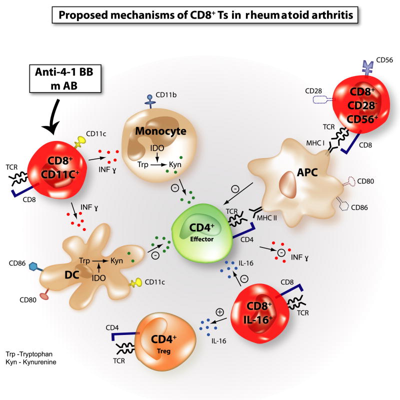
Rheumatologists have long been focused on developing novel immunotherapeutic agents to manage such prototypic autoimmune diseases as rheumatoid arthritis (RA) and systemic lupus erythematosus (SLE). The ultimate challenge in providing immunosuppressive treatment for patients with RA and SLE has derived from the dilemma that both protective and harmful immune responses result from adaptive immune responses, mediated by highly diverse, antigen-specific T and B cells endowed with powerful effector functions and the ability for long-lasting memory. As regulatory/suppressor T cells can suppress immunity against any antigen, including self-antigens, they emerge as an ideal therapeutic target. Several distinct subtypes of CD8(+) suppressor cells (Ts) have been described that could find application in treating RA or SLE. In a xenograft model of human synovium, CD8(+)CD28(-)CD56(+) T cells effectively suppressed rheumatoid inflammation. Underlying mechanisms involve conditioning of antigen presenting cells (APC). Adoptively transferred CD8(+) T cells characterized by IL-16 secretion have also exhibited disease-inhibitory effects. In mice with polyarthritis, CD8(+) Ts suppressed inflammation by IFNgamma-mediated modulation of the tryptophan metabolism in APC. In SLE animal models, CD8(+) Ts induced by a synthetic peptide exerted suppressive activity mainly via the TGFbeta-Foxp3-PD1 pathway. CD8(+) Ts induced by histone peptides were found to downregulate disease activity by secreting TGFbeta. In essence, disease-specific approaches may be necessary to identify CD8(+) Ts optimally suited to treat immune dysfunctions in different autoimmune syndromes.


Figures

Figure 1
Figure 1. The spectrum of phenotypic markers on the different subsets of CD8+ suppressor T cells (Ts)
Various cell surface markers, cytokines, and Foxp3 have been reported to be expressed by CD8+ Ts. Not all cell surface markers are expressed on one subset of CD8+ Ts. Not all cytokines are produced by one particular CD8+ Ts subset. Foxp3 expression was not determined in some CD8+ Ts.
Figure 2
Figure 2. Possible mechanisms of suppression by CD8+ suppressor T cells (Ts)
Various suppression mechanisms by CD8+ Ts have been proposed so far. (a) CD8+CD28Foxp3+ Ts upregulate inhibitory receptors, immunoglobulin-like transcript (ILT) 3 and ILT4 on antigen-presenting cells (APC), rendering APC tolerogenic. Costimulatory molecules on APC, CD80, and CD86 are also downregulated. (b) Immunosuppressive cytokines such as interleukin (IL)-10, TGFβ, IFNγ, and CCL4 are secreted by CD8+ Ts. (c) Major histocompatibility complex (MHC) class Ib molecule (Qa-1 in mice and HLA-E in humans)-restricted CD8+ Ts kill activated effector CD4+ T cells and dampen immune reactions. (d) CD8+ cytotoxic lymphocytes (CTL) work as suppressive T cells. When CTL encounter antigen-bearing APC, CTL may kill APC and attenuate immune responses.
Figure 3
Figure 3. Proposed mechanisms of CD8+ suppressor T cells (Ts) in rheumatoid arthritis (RA)
CD8+CD28CD56+ Ts transferred into SCID-chimera mice transplanted with synovial tissues from RA patients suppress the tissue inflammation measured by proinflammatory cytokines and chemokine expression, downmodulating the expression of costimulatory molecules CD80 and CD86 on antigen-presenting cells (APC) and synovial fibroblast-like cells (FLS). Interleukin (IL)-16-secreting CD8+ Ts suppress the tissue inflammation in the same SCID-chimera model. IL-16 is a natural ligand of the CD4 molecule and induces CD4+ T-cell anergy. IL-16 also may induce or recruit CD4+CD25+Foxp3+ Tregs in the tissue. In a mice collagen-induced arthritis model (CIA), CD8+CD11c+ Ts induced by the administration of anti-4-1 BB monoclonal antibodies (mAb) secrete IFNγ and induce indoleamine 2,3-dioxygenase (IDO) in CD11b+ monocytes and CD11c+ dendritic cells (DC). IDO catabolizes tryptophan (Trp) into its catabolite such as kynurenine (Kyn). Depletion of Trp and generated Kyn exert immunosuppressive effects on effector CD4+ T cells. Abbreviations: MHC = major histocompatibility complex; T-cell receptor = TCR.

References

    1. Gershon RK, Kondo K. Cell interactions in the induction of tolerance: the role of thymic lymphocytes. Immunology. 1970;18:723. - PMC - PubMed
    1. Sakaguchi S, Sakaguchi N, Asano M, Itoh M, Toda M. Immunologic self-tolerance maintained by activated T cells expressing IL-2 receptor alpha-chains (CD25). Breakdown of a single mechanism of self-tolerance causes various autoimmune diseases. J Immunol. 1995;155:1151. - PubMed
    1. Cools N, Ponsaerts P, Van Tendeloo VF, Berneman ZN. Regulatory T cells and human disease. Clin Dev Immunol. 2007;2007:89195. - PMC - PubMed
    1. Rouse BT. Regulatory T cells in health and disease. J Intern Med. 2007;262:78. - PubMed
    1. Valencia X, Lipsky PE. CD4+CD25+FoxP3+ regulatory T cells in autoimmune diseases. Nat Clin Pract Rheumatol. 2007;3:619. - PubMed

Publication types

MeSH terms