. 2008 Sep 22;8(1):163-72.
doi: 10.1102/1470-7330.2008.0026.

Pre-surgical high resolution ultrasound of facial basal cell carcinoma: correlation with histology

Affiliations

Pre-surgical high resolution ultrasound of facial basal cell carcinoma: correlation with histology

Francisco Bobadilla et al. Cancer Imaging. .

Abstract

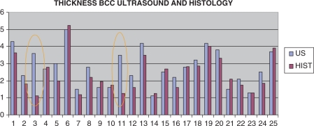
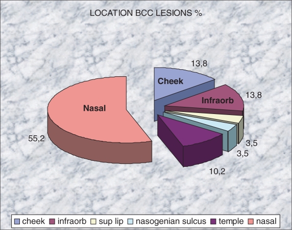
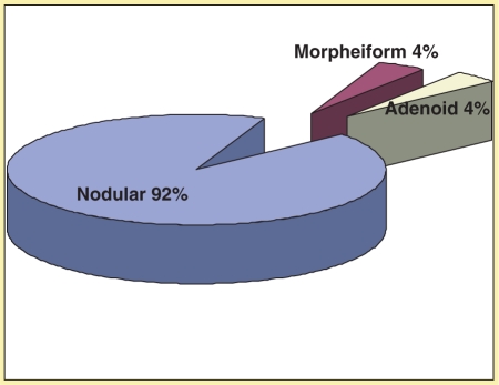
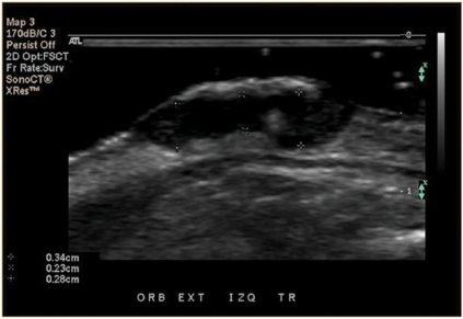
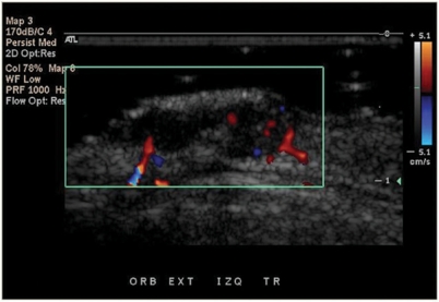
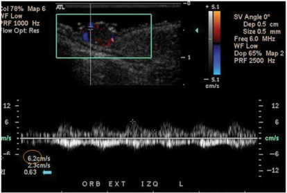
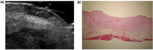
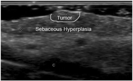
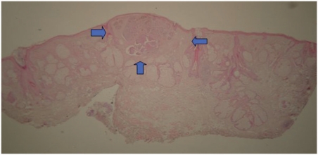
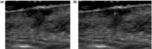
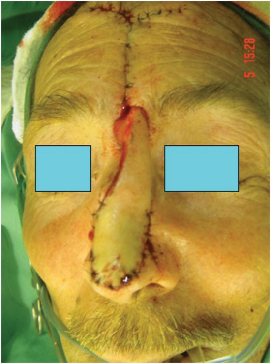
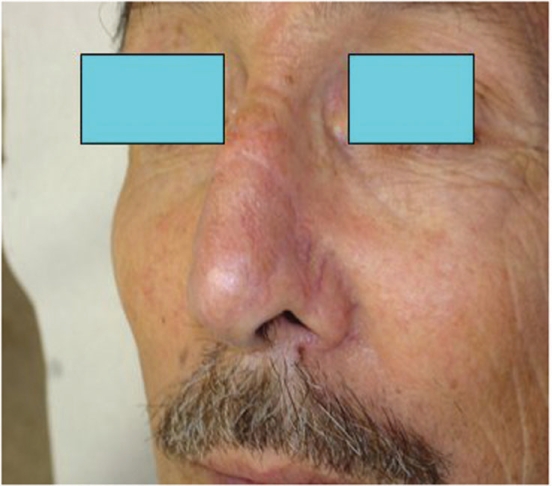
The aim of this study was to analyze the scope of pre-surgical high resolution ultrasound in basal cell carcinoma (BCC). BCC is the most common human cancer. According to recent large prospective studies incomplete excision of BCC is higher than expected. Pre-operative imaging may aid surgical planning by identifying the extent and location of a neoplasm, which can be interesting at zones with higher risk of recurrences such as the face. These are slow growing tumors but there are some aggressive types described that can involve deeper structures. Twenty-five patients were studied (10 F/15 M, 48-91 years old; mean age 69.5+/-11.5 years) with suspicious facial lesions < or =1.5 cm. Pre-surgical ultrasound with compact linear 15-7 MHz probe was performed. Ultrasound reported the morphology and thickness of the tumors. The results were correlated with the histology. Ultrasound identified 29 suspicious facial lesions that were removed with tumor-free borders at the first surgery and confirmed by histology. The main location was the nose. Two subclinical satellite lesions at the nasal zone were detected under ultrasound which led to a change in the surgery plan. The intraclass correlation coefficient (ICC) value was used to compare tumor thickness measurements between ultrasound and histology. ICC was considered as very good (0.9). Therefore, ultrasound can be useful to plan BCC surgery, it can recognize lesions, layers of involvement and vascularity patterns in a non-invasive way. It can show subclinical satellite lesions, even though the number of subclinical cases is small and require further investigations. It has a good thickness correlation with histology and may be used as a technique to monitor disease changes following non-invasive medical treatments in the future.


Figures

Figure 1
Figure 1
Compact linear probe, 15–7 MHz (variable frequency) used for the study.
Figure 2
Figure 2
Thickness (mm) correlation distribution between ultrasound and histology. Inside the orange circles are the two pitfall cases of the study.
Figure 3
Figure 3
Distribution of the location of the BCC lesions (%).
Figure 4
Figure 4
Histological distribution of the BCC lesions.
Figure 5
Figure 5
BCC tumor ultrasound shows an oval and hypoechoic lesion. Three thickness measurements (between calipers) demonstrate the irregularities at the deep borders that generate different distances. For the purpose of this study the largest distance was considered.
Figure 6
Figure 6
Color Doppler ultrasound shows the distribution of vessels (in red and blue) inside and at the deep portion of the tumor.
Figure 7
Figure 7
Spectral curve analysis color Doppler ultrasound of the arterial flow inside the tumor demonstrates the peak systolic velocity (circle) and the resistance index (arrow).
Figure 8
Figure 8
Ultrasound (a) and histology (b) of the same case showing a thickness difference of less than 1 mm (0.7 mm) between the methods and corresponding to a patient with a tumor measuring 4.2 mm thick by ultrasound and 3.5 mm by histology. Calipers are marking the lateral extension of the lesion, not the depth. Note the similar shape of the tumor under ultrasound and under 2 × zoom hematoxylin/eosin stain in histology.
Figure 9
Figure 9
Ultrasound of a case later described by histology as surrounded with sebaceous hyperplasia that overestimated the size of the tumor. Calipers show the measurements that were included in the ultrasound report. The (C) mark corresponds to the cartilage at the nasal ala. Notice the coarse, fuzzy and heterogeneous echoic pattern of the tissue, which differs from the lesions previously seen. Tumor and sebaceous hyperplasia zones are marked.
Figure 10
Figure 10
Histology of the same tumor as the patient in Fig. 9. The tumor is located between arrows and the rest was reported as sebaceous hyperplasia.
Figure 11
Figure 11
Ultrasound of a case described in histology as thickness overestimation because of the presence of a giant cell inflammatory reaction at the deepest portion. (a) Calipers show the actual measurements done by the ultrasound operator. In (b) the red lines show the angles formed by the inflammatory reaction at the profound part of the lesion. The tumor (T) was only located at the superficial area before the angles are formed.
Figure 12
Figure 12
Cutaneous frontal outflap surgery in a case with three BCC focuses on the nose. Two of the three focuses were subclinical and detected only at the ultrasound examination.
Figure 13
Figure 13
Post op result of the same patient as Fig. 9 after removal of all three BCC focuses in one time surgery.

Similar articles

Cited by

References

    1. Miller SJ. Biology of basal cell carcinoma (Part I) J Am Acad Dermatol. 1991;24:1–13. - PubMed
    1. Chuang TY, Popescu A, Su WP, Chute CG. Basal cell carcinoma: a population-based incidence study in Rochester, Minnesota. J Am Acad Dermatol. 1990;22:413–17. - PubMed
    1. Wong CS, Strange RC, Lear JT. Basal cell carcinoma. BMJ. 2003;327:794–8. - PMC - PubMed
    1. Strasswimmer J, Pierce MC, Park BH, Neel V, de Boer JF. Polarization-sensitive optical coherence tomography of invasive basal cell carcinoma. J Biomed Opt. 2004;9:292–8. - PubMed
    1. Preminger BA, Talmor M, Specht MC, Suzman M, Hoffman LA. The legacy of Icarus in the 21st century: report of a case of aggressive submental basal cell carcinoma resulting from frequent use of a metallic ultraviolet reflector. Ann Plast Surg. 2001;46:192–3. - PubMed

MeSH terms