. 2008 Sep 30;105(39):14755-62.
doi: 10.1073/pnas.0804216105. Epub 2008 Sep 23.

Navigating from hippocampus to parietal cortex

Affiliations

Navigating from hippocampus to parietal cortex

Jonathan R Whitlock et al. Proc Natl Acad Sci U S A. .

Abstract

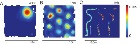
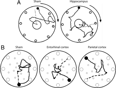
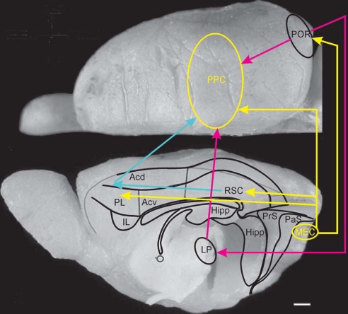
The navigational system of the mammalian cortex comprises a number of interacting brain regions. Grid cells in the medial entorhinal cortex and place cells in the hippocampus are thought to participate in the formation of a dynamic representation of the animal's current location, and these cells are presumably critical for storing the representation in memory. To traverse the environment, animals must be able to translate coordinate information from spatial maps in the entorhinal cortex and hippocampus into body-centered representations that can be used to direct locomotion. How this is done remains an enigma. We propose that the posterior parietal cortex is critical for this transformation.


Conflict of interest statement

The authors declare no conflict of interest.

Figures

Fig. 1.
Fig. 1.
Spatial firing properties of neurons in hippocampus (A; place cell), medial entorhinal cortex (MEC) (B; grid cell), and posterior parietal cortex (PPC) (C). In A and B, the rat runs freely in an open-field environment; in C, the rat traverses a complex maze with multiple segments and turns (Left, path of the animal; Center, rate map with lights on; Right, rate map in darkness). Firing rates are color-coded, with red showing maximal rate and blue minimum (scale bar to the right). Note that the parietal neuron fires at specific epochs along the trajectory and that firing is independent of visual inputs. [C is adapted with permission from ref. (Copyright 2006, Neuron).]
Fig. 2.
Fig. 2.
Disrupted path integration in rats with lesions of hippocampus (A), entorhinal cortex, or PPC (B). The rats were trained to leave a starting refuge, enter a large circular open field, search for a large food pellet, and bring the food pellet back to the refuge. The start box was placed beneath one of eight holes along the periphery of the circle (small filled circle). In A, the arena was slowly rotated, at subvestibular speed, as the rat searched for food. Gray circles indicate position of the start box after rotation. In B, there was no rotation. Outward paths are shown as solid lines in both A and B; return paths are shown as dashed lines (only from the food location to the first choice). Control animals generally chose the shortest path back to the point where they started, whereas rats with lesions in hippocampus, entorhinal cortex, or PPC generally chose the wrong hole, indicating that path integration was disrupted. Note that, although the lesioned rat in A ran directly to the novel target location, the dispersion of first choices was large in the hippocampal group as a whole. [Adapted with permission from ref. (Copyright 2004, Hippocampus) and ref. (Copyright 2004, Experimental Brain Research).]
Fig. 3.
Fig. 3.
Dorsal view of the right hemisphere of the rat brain showing the different delineations of the parietal cortex of the rat as reported by Krieg (100), in red; Palomero-Gallagher and Zilles (108), in yellow; Miller and Vogt (109), in magenta; and Burwell and Amaral (67), in blue. Whether or not to include parts of Oc2 in the PPC (i.e., the lateromedial extent of the PPC, or area 7) is still controversial. The figures are based on the original diagrams but have been redrawn onto a standard representation. (Scale bar, 1 mm.)
Fig. 4.
Fig. 4.
Predictive relationship between firing in monkey PPC and the next movement to be made in a choice task. The monkey was trained to indicate the location of a target on a screen by using either an eye movement (saccade, when the cue was red) or an arm movement (reach, when the cue was green). Firing rates are shown for two cells as a function of time, including onset and offset of the cue and initiation of the response (Go). (Top) Raster plots; (Bottom) smoothed peristimulus time histograms. The cell in A is from the lateral intraparietal area; the cell in B is from the parietal reach region. When the cue was on, firing was similar in the two regions. When the cue was turned off, different patterns emerged. Saccades were preceded by preferential firing in the lateral intraparietal area; reaches were preceded by stronger firing in the parietal reach region. Thus, firing during the delay period depends on the motor output required to solve the task, suggesting that the PPC represents intended movements to specific locations. [Reproduced with permission from ref. (Copyright 2007, Neuron).]
Fig. 5.
Fig. 5.
Dorsal and midsagittal view of the rat brain indicating the proposed routes by which information from the medial entorhinal grid cell area may reach PPC. The most likely route by way of the connections mediated by the postrhinal cortex is indicated (yellow and purple) as well as the multisynaptic routes using pathways through the lateral posterior complex of the thalamus (purple), and the retrosplenial cortex (RSC) and dorsal portion of medial prefrontal cortex (PL and ACd) (yellow and blue). (Scale bar, 1 mm.)

References

    1. O'Keefe J, Nadel L. The Hippocampus as a Cognitive Map. Oxford: Clarendon; 1978.
    1. Biegler R. Possible uses of path integration in animal navigation. Anim Learn Behav. 2000;28:257–277.
    1. Etienne AS, Jeffery KJ. Path integration in mammals. Hippocampus. 2004;14:180–192. - PubMed
    1. O'Keefe J, Dostrovsky J. The hippocampus as a spatial map: Preliminary evidence from unit activity in the freely-moving rat. Brain Res. 1971;34:171.175. - PubMed
    1. O'Keefe J. Place units in the hippocampus of the freely moving rat. Exp Neurol. 1976;51:78–109. - PubMed