. 2008 Sep 23;105(38):14336-41.
doi: 10.1073/pnas.0805942105.

Isolation of human monoclonal antibodies by mammalian cell display

Affiliations

Isolation of human monoclonal antibodies by mammalian cell display

Roger R Beerli et al. Proc Natl Acad Sci U S A. .

Abstract

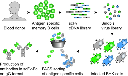
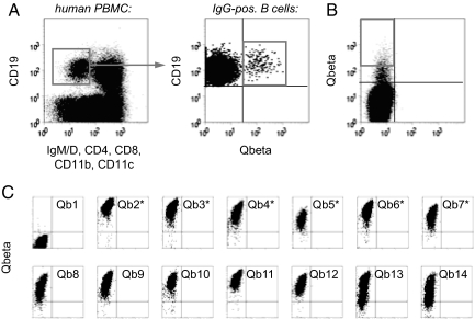
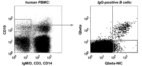
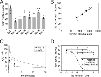
Due to their low immunogenicity in patients, humanized or fully human mAbs are becoming increasingly important for the treatment of a growing number of diseases, including cancer, infections, and immune disorders. Here, we describe a technology allowing for the rapid isolation of fully human mAbs. In contrast to previously described methods, B cells specific for an antigen of interest are directly isolated from peripheral blood mononuclear cells (PBMC) of human donors. Recombinant, antigen-specific single-chain Fv (scFv) libraries are generated from this pool of B cells and screened by mammalian cell surface display by using a Sindbis virus expression system. This method allows isolating antigen-specific antibodies by a single round of FACS. The variable regions (VRs) of the heavy chains (HCs) and light chains (LCs) are isolated from positive clones and recombinant fully human antibodies produced as whole IgG or Fab fragments. In this manner, several hypermutated high-affinity antibodies binding the Qbeta virus like particle (VLP), a model viral antigen, as well as antibodies specific for nicotine were isolated. All antibodies showed high expression levels in cell culture. The human nicotine-specific mAbs were validated preclinically in a mouse model. Thus, the technology presented here allows for rapid isolation of high-affinity, fully human antibodies with therapeutic potential from human volunteers.


Conflict of interest statement

Conflict of interest statement: All authors are employees and owners of stock options of Cytos Biotechnology AG, and therefore declare potential competing interest.

Figures

Fig. 1.
Fig. 1.
Isolation of human antibodies by mammalian cell display. Antigen-specific, isotype switched B cells are isolated from the PBMC of a human donor by FACS by using fluorescently labeled antigen as a bait. RNA isolated from these specific B cells is used to generate a random combinatorial scFv library. This enriched library, typically consisting of high-affinity binders (functional pairing of HCVR and LCVR, green), low affinity binders (suboptimal pairing, blue), and nonbinders (nonfunctional pairing, gray), is then converted to a high-titer Sindbis virus expression library. Infection of BHK cells at a low MOI generates a pool of infected cells, each expressing at their surface one specific antibody. Single cells expressing a functional antibody are then isolated by flow cytometry and sorted onto a monolayer of BHK cells. Once the virus has spread and expression of antigen-specific antibody is verified, the VRs are cloned and the antibody can be expressed in any desired format.
Fig. 2.
Fig. 2.
Screening for Qβ-specific antibodies. (A) Isolation of Qβ-specific B cells by FACS. Human PBMC were stained for the presence or absence of the indicated surface markers and analyzed for expression of Qβ-specific antibodies. IgM-IgD-negative, isotype switched B cells (as gated in Left) specifically binding to Qβ (as gated in Right) were sorted for library construction. (B) Sorting of BHK cells displaying Qβ-specific antibodies. BHK cells were infected at a low MOI with a Sindbis virus library encoding scFv antibodies constructed from Qβ-specific B cells. After staining with Alexa 647 nm-labeled Qβ, cells displaying specific antibodies were sorted onto semiconfluent BHK feeder cells (1 cell per well). (C) Rescreening for Qβ-specific antibodies. Two to three days after sorting, BHK cells showing signs of viral infection were analyzed for binding to Qβ. The first 14 of the 276 wells analyzed are shown. Antibodies expressed by those clones indicated with an asterisk were selected for gene rescue and sequencing.
Fig. 3.
Fig. 3.
Isolation of nicotine-specific B cells by FACS. Human PBMC were stained for the presence or absence of the indicated surface markers and analyzed for expression of Qβ- and nicotine-specific antibodies. Isotype switched B cells (as gated in Left) specifically binding to nicotine but not Qβ (as gated in Right) were sorted for library construction.
Fig. 4.
Fig. 4.
Characterization of nicotine-specific antibodies. (A) Inhibition of nicotine entry into brain. Mice were injected i.p. with 0.5 mg of the indicated human IgG2. One day later, mice were injected i.v. with 750 ng of tritium-labeled nicotine. Mice were killed after 5 min and nicotine concentrations in brains were measured. Individual mice are shown; bars indicate average values. IgG, control human IgG. *, P < 0.001 vs. IgG; **, P = 0.003 vs. IgG. (B) Dose-response of inhibition of nicotine entry into brain by IgG2-Nic12. Mice were injected i.p. with 0.1, 0.3, 1, or 3 mg of mAb-Nic12. One day later, mice were injected i.v. with 750 ng of tritium-labeled nicotine. Mice were killed after 5 min and nicotine concentrations in brains were measured. Antibody concentrations in sera were determined and plotted against the percentage nicotine-reduction in the brain. (C) Time course of inhibition of nicotine entry into brain by IgG2-Nic12. Mice were injected with 0.5 mg antibody, challenged with 750 ng of tritium-labeled nicotine and nicotine in the brain was measured at the indicated times. Averages of 4 to 5 mice are given with standard deviations. IgG, control human IgG. *, P < 0.001 vs. IgG. (D) Specificity of mAb-Nic12. The binding of tritium-labeled (−)-nicotine (56 nM) to mAb-Nic12 (50 nM) was measured by equilibrium dialysis in the presence of increasing concentrations of unlabeled (−)-nicotine, (−)-cotinine, or acetylcholine. Averages of two independent experiments are given with standard deviations.

References

    1. Reichert JM, Rosensweig CJ, Faden LB, Dewitz MC. Monoclonal antibody successes in the clinic. Nat Biotechnol. 2005;23:1073–1078. - PubMed
    1. Miller RA, Oseroff AR, Stratte PT, Levy R. Monoclonal antibody therapeutic trials in seven patients with T-cell lymphoma. Blood. 1983;62:988–995. - PubMed
    1. Ratner B. Allergy, Anaphylaxis and Immunotherapy, Basic Principles and Practice. Baltimore: Williams and Wilkins; 1943.
    1. Foote J, Winter G. Antibody framework residues affecting the conformation of the hypervariable loops. J Mol Biol. 1992;224:487–499. - PubMed
    1. Riechmann L, Clark M, Waldmann H, Winter G. Reshaping human antibodies for therapy. Nature. 1988;332:323–327. - PubMed

MeSH terms

LinkOut - more resources