. 2008 Dec;74(12):1557-67.
doi: 10.1038/ki.2008.412. Epub 2008 Aug 27.

Effects of the renal medullary pH and ionic environment on vasopressin binding and signaling

Affiliations

Effects of the renal medullary pH and ionic environment on vasopressin binding and signaling

Elena A Zalyapin et al. Kidney Int. 2008 Dec.

Abstract

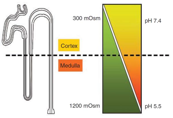
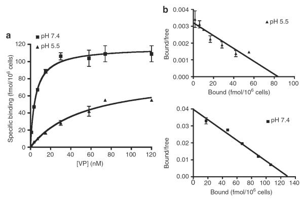
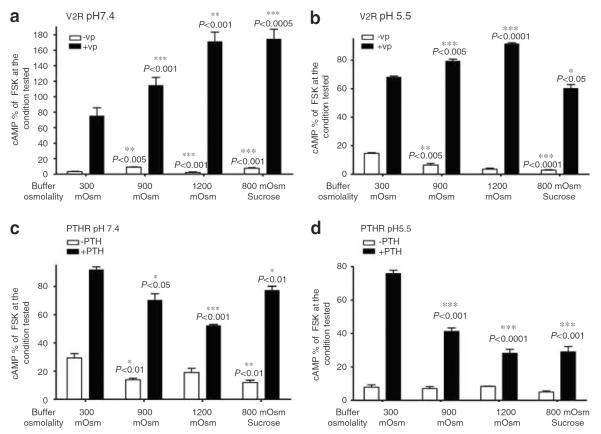
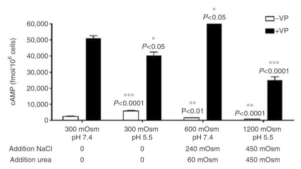
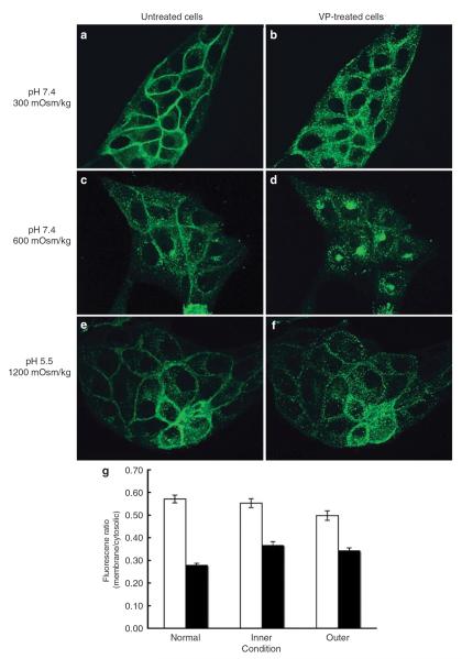
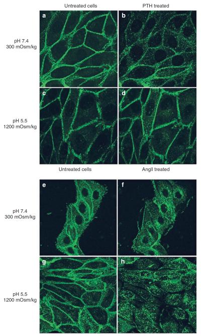
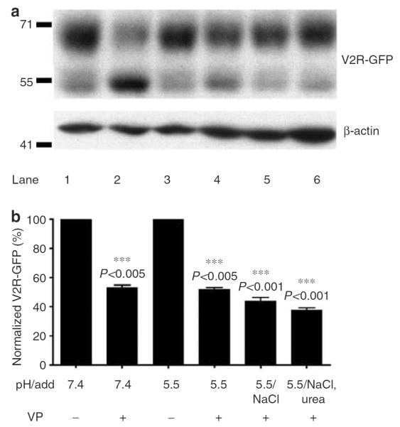
The kidney has a cortico-medullary interstitial gradient of decreasing pH and increasing concentrations of sodium chloride and urea, but the influence of these gradients on receptor signaling is largely unknown. Here, we measured G-protein coupled receptor function in LLC-PK1 cells acutely exposed to conditions mimicking different kidney regions. Signaling through the parathyroid hormone receptor, normally expressed in the cortex, was greatly reduced at an acidic pH similar to that of the inner medulla. Parathyroid hormone receptor, tagged with green fluorescent protein, showed no ligand-induced internalization. In contrast, under both acidic and hyperosmotic conditions, vasopressin increased intracellular cAMP, and upon binding to its type 2 receptor (V2R) was internalized and degraded. Dose-displacement binding assays with selective vasopressin/oxytocin receptor ligands under inner medullary conditions indicated a shift in the V2R pharmacological profile. Oxytocin did not bind to the V2R, as it does under normal conditions and the vasopressin type 1 receptor (V1R) had reduced affinity for vasopressin compared to the V2R in low pH and high osmolality. We suggest that the cortico-medullary gradient causes a receptor-specific selectivity in ligand binding that is of functional significance to the kidney. While the gradient is important for urinary concentration, it may also play a substantial role in fine-tuning of the vasopressin response through the V2R.


Figures

Figure 1
Figure 1. The collecting duct is exposed to a corticomedullary gradient
The kidney collecting duct is exposed to a corticomedullary gradient of decreasing pH and increasing concentrations of NaCl and urea.
Figure 2
Figure 2. Under conditions of acidic pH the affinity of VP for V2R is lower than at neutral pH
LLC-FLAG-V2R cells were grown to confluence following incubation with different concentrations of [3H]VP at pH 7.4 (■) and pH 5.5 (▲) for 3 h at 4°C (a). Scatchard plot representation of binding saturation experiments at pH 7.4 (■) and pH 5.5 (▲) (b). Means of triplicate values are shown, representative of data from four independent experiments (mean±s.e.m.).
Figure 3
Figure 3. Hypertonicity increases cAMP signaling of VP-stimulated V2R, but decreases signaling of PTH stimulated PTHR
cAMP accumulation was tested using a cAMP assay as described in section `Materials and Methods'. cAMP accumulation was studied in the presence or absence of 1 μm VP or PTH (open bars and closed bars, respectively). VP was used to stimulate LLC-FLAG-V2R and PTH was used to stimulate LLC-PTHR cells at pH 7.4 (a, c) or at pH 5.5 (b, d). Experiments were performed in triplicate. The number of experiments performed is shown in parenthesis. Data are shown as mean±s.e.m. Maximal forskolin response at pH 7.4, 300 mOsm for LLC-FLAG-V2R is 11,234±135 fmol/106 cells. Maximal forskolin response at pH 7.4, 300 mOsm for LLC-PTHR is 12,429±491 fmol/106 cells. We compared by t-test the amount of cAMP before VP stimulation at pH 7.4, 300 mOsm to other nonstimulated conditions. Intracellular cAMP accumulation following VP stimulation at pH 7.4, 300 mOsm was compared with VP-induced cAMP accumulation occurring under all other conditions. Significance of t-test is indicated by asterisks.
Figure 4
Figure 4. cAMP accumulation following V2R stimulation by VP occurs under `harsh' conditions mimicking those of the inner medulla
cAMP accumulation was studied in the presence or absence of 1 μm VP (open bars and closed bars, respectively). VP was diluted in normal and acidic buffer in the presence of increasing amounts of sodium and urea. Experiments shown represent an average of three different experiments performed in triplicate (mean±s.e.m.). Maximal forskolin response at pH 7.4, 300 mOsm is 12, 270±467 fmol/106 cells.
Figure 5
Figure 5. V2R internalizes following VP stimulation under `harsh' conditions mimicking those of the inner medulla
LLC-V2R-GFP cells were incubated in the absence (a, c, and e) or the presence of 1 μm VP (b, d, and f) under conditions mimicking normal (upper panel), outer (middle panel), and inner medulla (lower panel). In (d), a perinuclear accumulation of V2R was observed after internalization. This phenomenon is currently under more detailed investigation. These results are representative of three independent assays. Three other assays were performed and analyzed by epifluorescence microscopy. Quantification of membrane fluorescence was analyzed using IPLab software (g) as described in section `Materials and Methods'; mean±s.e.m.
Figure 6
Figure 6. AT1R receptors internalize following AngII stimulation under `harsh' inner medullary conditions, whereas PTH-stimulated PTHR does not
LLC-PTHR-GFP (a–d) and LLC-AT1R-GFP (e–h) cells, stably expressing either PTHR-GFP or AT1R-GFP were incubated in the absence (a, c and e, g) or presence of 1 μm of their respective agonists (b, d and f, h). Cells were incubated with either neutral (a, b and e, f) or acidic hyperosmotic medium (c, d and g, h). Sequential images were acquired using spinning disk confocal microscopy. Acquired images of cells taken before and after 30 min of agonist stimulation are representative of four independent experiments.
Figure 7
Figure 7. VP stimulated V2R-GFP degrades under conditions of both acidic and neutral pH
V2R-GFP degradation after 4 hours of VP stimulation (at 37 °C) under both neutral and acidic conditions was studied by Western blot analysis (a). LLC-V2R-GFP cells were incubated in the absence (lanes 1 and 3) or presence (lanes 2 and 4) of VP. In the absence of VP, 71 and 55 kDa bands are observed corresponding to intact, glycosylated V2R and to a degraded fragment of this receptor, respectively. The presence of VP reduced the intensity of the 71 kDa band (lane 2). Similar results were observed under acidic conditions (lanes 4–6). Disappearance of the upper band intensity was quantified in relation to the intensity of corresponding β-actin bands, used as a loading control (b). This is a representative blot from three independent experiments; Mean±SD. ***P>0.005 between VP treated and untreated conditions, ***P>0.001 between sample incubated at pH 5 in the presence of VP and in the presence of either NaCl or Urea.

References

    1. Birnbaumer M, Seibold A, Gilbert S, et al. Molecular cloning of the receptor for human antidiuretic hormone. Nature. 1992;357:333–335. - PubMed
    1. Lolait SJ, O'Carroll AM, McBride OW, et al. Cloning and characterization of a vasopressin V2 receptor and possible link to nephrogenic diabetes insipidus. Nature. 1992;357:336–339. - PubMed
    1. Morel A, O'Carroll AM, Brownstein MJ, et al. Molecular cloning and expression of a rat V1a arginine vasopressin receptor. Nature. 1992;356:523–526. - PubMed
    1. Sugimoto T, Saito M, Mochizuki S, et al. Molecular cloning and functional expression of a cDNA encoding the human V1b vasopressin receptor. J Biol Chem. 1994;269:27088–27092. - PubMed
    1. Calo G, Rizzi A, Traina L, et al. Pharmacological characterization of a vasopressin V1 receptor in the isolated human gastric artery. Life Sci. 1997;60:PL63–PL68. - PubMed

Publication types

LinkOut - more resources