. 2008 Sep 24;3(9):e3271.
doi: 10.1371/journal.pone.0003271.

Activin A induces Langerhans cell differentiation in vitro and in human skin explants

Affiliations

Activin A induces Langerhans cell differentiation in vitro and in human skin explants

Tiziana Musso et al. PLoS One. .

Abstract

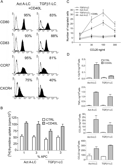
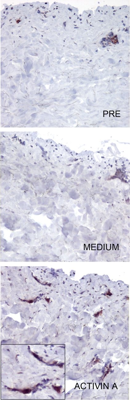
Langerhans cells (LC) represent a well characterized subset of dendritic cells located in the epidermis of skin and mucosae. In vivo, they originate from resident and blood-borne precursors in the presence of keratinocyte-derived TGFbeta. In vitro, LC can be generated from monocytes in the presence of GM-CSF, IL-4 and TGFbeta. However, the signals that induce LC during an inflammatory reaction are not fully investigated. Here we report that Activin A, a TGFbeta family member induced by pro-inflammatory cytokines and involved in skin morphogenesis and wound healing, induces the differentiation of human monocytes into LC in the absence of TGFbeta. Activin A-induced LC are Langerin+, Birbeck granules+, E-cadherin+, CLA+ and CCR6+ and possess typical APC functions. In human skin explants, intradermal injection of Activin A increased the number of CD1a+ and Langerin+ cells in both the epidermis and dermis by promoting the differentiation of resident precursor cells. High levels of Activin A were present in the upper epidermal layers and in the dermis of Lichen Planus biopsies in association with a marked infiltration of CD1a+ and Langerin+ cells. This study reports that Activin A induces the differentiation of circulating CD14+ cells into LC. Since Activin A is abundantly produced during inflammatory conditions which are also characterized by increased numbers of LC, we propose that this cytokine represents a new pathway, alternative to TGFbeta, responsible for LC differentiation during inflammatory/autoimmune conditions.


Conflict of interest statement

Competing Interests: The authors have declared that no competing interests exist.

Figures

Figure 1
Figure 1. Activin A promotes Langerhans cell differentiation from human CD14+ monocytes.
(A) Phenotypic analysis of monocytes cultured for 6 days with GM-CSF and IL-4 in the presence of Activin A (Act A-LC) or TGFβ1 (TGFβ1-LC). Cells were stained with the indicated moAbs (filled histograms) or isotype-matched negative control moAbs (open histograms). Percentages of positive cells are shown in the upper right corner of each histogram. The figure shows one experiment representative of at least five independent cultures. (B) Electron microscopy analysis of Act A-LC. Act A-LC exhibited abundant dendritic membrane protrusions and lobulated or indented nuclei (left panel, 3,000X, bar 40 µm). Cytoplasm presented a rough endoplasmic reticulum, many multilamellar organelles and numerous electron-dense structures reminiscent of Birbeck granules (right panel, 12,000X, bar 1 µm). The inset shows rod-shaped Birbeck granules (200,000X, bar 20 µm). (C) TGFβ1 and Activin A mRNA expression in Act A-LC and TGFβ1-LC cultures. Monocytes were cultured in the presence of Act A or TGFβ1 for the indicated time and the expression of TGFβ1 and Activin A mRNA was determined by real-time PCR, relative to GAPDH mRNA used as internal control. The expression level in freshly isolated monocytes was assumed as the 1.0 value. Similar results were obtained in three different donors. (D) Effects of different TGF family members on LC differentiation. Monocytes were cultured for 6 days with GM-CSF in the presence of 10 ng/ml TGFβ1, 100 ng/ml Activin A, or 100 ng/ml BMP6 and analyzed for Langerin, E-caderin and CCR6 expression by flow cytometry analysis. Data are representative of at least four independent cultures.
Figure 2
Figure 2. Phenotypical and functional characterization of CD40L-activated Act A-LC
(A) Expression of maturation markers by Act A-LC. Act A-LC were incubated with CD40L-transfected fibroblasts for 40 hrs and stained with anti-CD80, CD83, CCR7 and CXCR4 moAbs (filled histograms) or isotype-matched negative control Abs (open histograms). Results obtained with TGFβ1-LC are also shown for comparison. The percentage of positive cells is reported in each panel. Data shown are representative of three independent experiments. (B) Allostimulatory capacity of Act A-LC. Irradiated immature or CD40L-matured Act A-LC (or TGFβ1-LC) were cultured with 2×10⁵ allogeneic purified T cells. Proliferation was assayed as uptake of [H3]thymidine added in the last 16 hrs of a 6-day culture assay. Results are expressed as mean counts per minute (cpm)±SD of one representative experiment performed in triplicate. Values are at the net of T cell proliferation in the absence of DC (3250±250 cpm). (C) Act A-LC migrate in response to CCL20. Immature or CD40L-mature Act A-LC or TGFβ1-LC were applied to the upper wells of the chemotaxis chamber. CCL20 was added to the lower level of the chamber. The number of cells migrated to the lower chamber was counted. Each assay was performed in triplicate and the results are expressed as the mean±SD number of migrated cells (representative of three experiments). (D) Cytokine release by Act A-LC. Immature or CD40L-mature Act A-LC or TGFβ1-LC were assessed for their ability to release the indicated cytokines by ELISA. Results are the average determination (±SD) of four independent experiments.
Figure 3
Figure 3. Intradermal injection of Activin A induces the differentiation of dermal and epidermal Langerhans cells in human skin explants.
Langerin expression was evaluated in the epidermis and dermis (full thickness skin explants) of skin explants, untreated and 72 hrs after i.d. injection of medium or 100 ng Activin A (magnification 100X, inset 400X) The number of Langerin+ cells were quantified in skin explants by evaluating six different skin sections (0.05 mm2/field; means±SD). * p<0.05 by Student's t test vs. medium (lower right panel).
Figure 4
Figure 4. Activin A induces Langerhans cells differentiation in epidermis-depleted skin explants.
Langerin expression was evaluated in the dermal layer, separated from skin explants by dispase digestion and subsequently treated for 72 hrs after i.d. injection with 100 ng Activin A (magnification 100X, inset 400X.).
Figure 5
Figure 5. Dermal accumulation of Langerhans cells in lichen planus is associated to abundant production of Activin A.
Sections from normal skin (NS) (a and d) and lichen planus (LP) (b, c, e, f) biopsies were stained for Langerin (ac) and Activin A (d–f). In normal skin, Langerin+ cells are regularly distributed in basal and suprabasal layers and show multiple fine dendrites; no positive cells are detectable in the dermis (panel a). In LP biopsies, in addition to intraepidermal LC, accumulation of Langerin+ cells is observed in the dermis within the dense monuclear cell infiltrate (panel b). At high power view, Langerin+ cells show an ovoidal/dendritic shape (panel c) and are found surrounding Factor VIII+ dermal blood vessels (arrow head, inset in c). Serial sections from the same tissue blocks were stained for Activin A. Normal skin (panel d) showed weak intraepithelial reactivity (red arrow head); in the dermis, mast cells and occasional spindle cells were positive for Activin A (black arrow heads). In LP, Activin A was strongly induced in the superficial layers of epidermis; in the dermis, a diffuse reactivity can be observed in numerous cells within the inflammatory infiltrate (panel e). This cell population includes endothelial cells and a mixture of non-lymphoid mononuclear cells (panel f). Magnification 100x (a, b, d, e; scale bar 200 micron) and 400x (c, f; scale bar 50 micron).

References

    1. Banchereau J, Briere F, Caux C, Davoust J, Lebecque S, et al. Immunobiology of dendritic cells. Annu Rev Immunol. 2000;18:767–811. - PubMed
    1. Bell D, Young JW, Banchereau J. Dendritic cells. Adv Immunol. 1999;72:255–324. - PubMed
    1. Steinman RM, Banchereau J. Taking dendritic cells into medicine. Nature. 2007;449:419–426. - PubMed
    1. Stingl G, Katz SI, Shevach EM, Rosenthal AS, Green I. Analogous functions of macrophages and Langerhans cells in the initiation in the immune response. J Invest Dermatol. 1978;71:59–64. - PubMed
    1. Strobl H, Riedl E, Bello-Fernandez C, Knapp W. Epidermal Langerhans cell development and differentiation. Immunobiology. 1998;198:588–605. - PubMed

Publication types

MeSH terms