. 2008 Oct;10(10):1138-45.
doi: 10.1593/neo.08430.

Hsf1 is required for the nuclear translocation of p53 tumor suppressor

Affiliations
Free PMC article

Hsf1 is required for the nuclear translocation of p53 tumor suppressor

Qiang Li et al. Neoplasia. 2008 Oct.
Free PMC article

Abstract

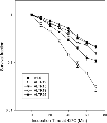
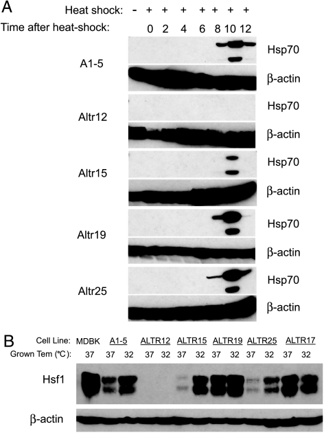
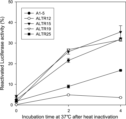
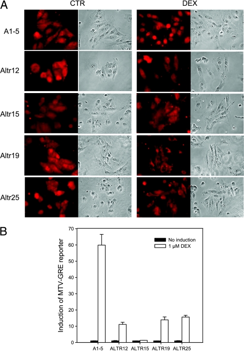
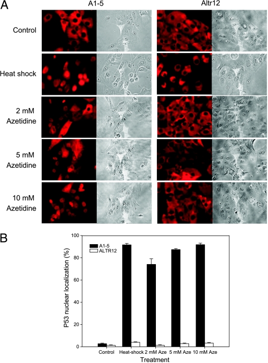
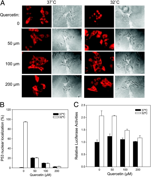
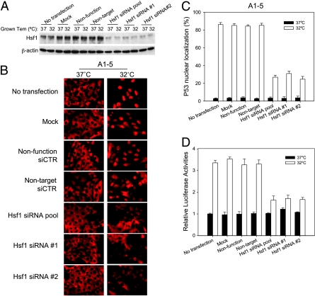
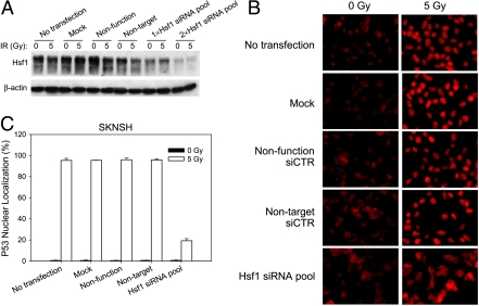
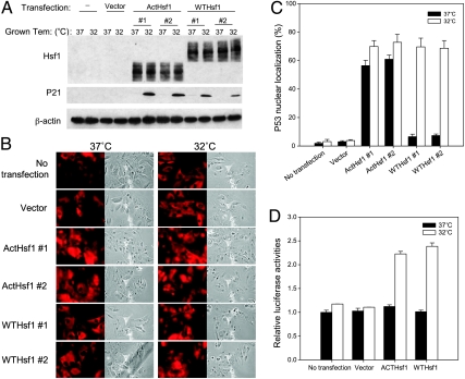
Although the p53 tumor suppressor is most frequently inactivated by genetic mutations, exclusion from the nucleus is also seen in human tumors. We have begun to examine p53 nuclear importation by isolating a series of mutant cells in which the temperature-sensitive murine p53(Val135) mutant is sequestered in the cytoplasm. We previously showed that that three of them (ALTR12, ALTR19, and ALTR25) constituted a single complementation group. Here, we found that ALTR12 cells are more sensitive to heat stress than either ALTR19 or ALTR25 and that there was a complete lack of induction of Hsp70 in response to heat shock. Western blot analysis showed no expression of the Hsf1 transcription factor, and neither heat shock nor azetidine could induce p53 nuclear localization in ALTR12 cells but did in parental A1-5 cells. Suppression of Hsf1 in A1-5 cells with quercetin or an Hsf1 siRNA reduced p53 nuclear importation and inhibited p53-mediated activation of a p21 reporter. Most convincingly, p53 nuclear importation could be restored in ALTR12 cells by introducing an exogenous Hsf1 gene. Collectively, our result suggests that Hsf1 is required for p53 nuclear importation and activation and implies that heat shock factors play a role in the regulation of p53.


Figures

Figure 1
Figure 1
Heat shock survival curve of A1–5 and ALTR cells. Cells (80% confluence) were heat-shocked and plated as described in the Materials and Methods section. After incubation at 37°C for ∼10 days, colonies were stained with crystal violet and read on a colony counter. Graphs show the mean surviving fraction ± SE and are from three independent experiments.
Figure 2
Figure 2
Expression of Hsp70 and HSF1 is impaired in ALTR12 cells. (A) Induction of Hsp70 in A1–5 and ALTR cells was examined by Western blot analysis. Cells at 80% confluence were heat-shocked for 20 minutes at 42°C. After incubation at 37°C for different time points, cells were harvested and analyzed by Western blot analysis using an anti-Hsp70 antibody. (B) The expression of Hsf1 in A1–5 and ALTR cells was examined by Western blot analysis. Confluent A1–5 and ALTR cells grown at 37°C or 32°C were harvested and analyzed by Western blot analysis using an anti-Hsf1 antibody.
Figure 3
Figure 3
Luciferase refolding is defective in ALTR12 cells. A1–5 and ALTR cells were transfected with pSa244 and incubated for 48 hours to allow for optimal luciferase expression. Cells were then exposed to 42°C for 20 minutes in the presence of protein synthesis inhibitor cycloheximide (10 µg/ml). Luciferase activity was determined either immediately after heating or after a 2- or 4-hour recovery period at 37°C. Graphs show the mean ± SE from three independent experiments.
Figure 4
Figure 4
Nuclear localization of the GR was inhibited in ALTR cells. (A) A1–5 and ALTR cells were grown on coverslips. After treatment with or without DEX for 3 hours, GR localization were stained using an anti-GR antibody. Photomicrographs of a typical experiment are shown. (B) A1–5 and ALTR cells that were transfected with either the pMMfluc, which encodes a firefly luciferase reporter under the control of MTV-GRE promoter, or the pRenilla-Luc as internal control. At 24 hours after transfection, cells were treated with or without DEX, and luciferase activities were measured after 6 hours. Luciferase activities at any particular treatment were corrected for variations in transfection efficiency using the internal control. Graphs show the mean ± SE from three independent experiments.
Figure 5
Figure 5
Heat shock-inducible p53 nuclear import is inhibited in ALTR12 cells. (A) A1–5 or ALTR12 cells were treated with heat shock (42°C for 20 minutes) or azetidine at the concentration indicated. After incubation at 37°C for 3 hours, cells were stained for p53 localization using PAb421 antibody. The photomicrographs shown represent a typical result. (B) The fraction of nuclear localized p53 in A was quantified. Graphs show the mean ± SE from three independent experiments.
Figure 6
Figure 6
Quercetin suppressed p53 nuclear localization in A1–5 cells. (A) A1–5 cells were grown on coverslips at 37°C to 60% confluence. The cells were then treated with different concentrations of quercetin or left untreated for 2 hours at 37°C. The cells were then shifted to 32°C for 5 hours and then stained for p53 localization by PAb421 antibody. The photomicrographs shown represent a typical result. (B) The fraction of nuclear localized p53 in A was quantified. Graphs show the mean ± SE from three independent experiments. (C) A1–5 cells stably transfected with pWWP-Luc-Neo plasmid, which encodes a firefly luciferase under the control of p21 promoter, were grown at 37°C to 80% confluence. After treatment with different concentrations of quercetin for 2 hours at 37°C, the cells were shifted to a 32°C incubator for 6 hours and then assayed for luciferase activity. Graphs show the mean ± SE from three independent experiments.
Figure 7
Figure 7
Suppression of Hsf1 with siRNA inhibits p53 nuclear localization. (A) A1–5 cells were transfected with the indicated siRNA at 37°C. Forty-eight hours later, either the cells were harvested and extracts were prepared (37°C) or the incubation temperature was shifted to 32°C for 5 hours before preparing extracts (32°C). Levels of Hsf1 were determined by immunoblot analysis with the appropriate antibody. β-Actin levels were used as loading controls. (B) A1–5 cells were grown on coverslips at 37°C to 60% confluence and then transfected with the indicated siRNA. Forty-eight hours later, the coverslips were either immediately stained for p53 localization (37°C) or shifted to 32°C for 5 hours and then stained for p53 localization by PAb421. (C) The fraction of cells with nuclear localized p53 in panel (B) was quantified. Graphs show the mean ± SE from three independent experiments. (D) A1–5 cells stably transfected with pWWP-Luc-Neo plasmid were grown at 37°C to 60% confluence. Forty-eight hours after transfection with the indicated siRNA, either the cells were harvested immediately and assayed for luciferase activity (37°C) or the incubation temperature was shifted to 32°C for an additional 6 hours and then assayed for luciferase activity (32°C). Graphs show the mean ± SE from three independent experiments.
Figure 8
Figure 8
Hsf1 knockdown in SK-N-SH cells suppresses p53 nuclear localization. (A) SK-N-SH cells were grown to 60% confluence and then transfected with the indicated siRNA either once (1x) or twice with a 24-hour interval (2x). Forty-eight hours after the last transfection of siRNA, the transfected cells were either treated with a 5-Gy ionizing radiation or left untreated. Cells were harvested, and extracts were prepared 3 hours after irradiation. Hsf1 expression was determined by Western blot analysis using an anti-Hsf1 antibody. β-Actin levels are depicted as the loading control. (B) SK-N-SH cells were grown on coverslips at 37°C to 60% confluence and were transfected with the indicated siRNA. The cells were transfected twice with the Hsf1 siRNA as in panel (A). Forty-eight hours later, the cells were either treated with a 5-Gy ionizing radiation or left untreated. p53 localization was stained using DO1 antibody 3 hours after irradiation. (C) The fraction of cells with nuclear localized p53 in panel (B) was quantified. Graphs show the mean ± SE from three independent experiments.
Figure 9
Figure 9
Exogenous Hsf1 restores p53 nuclear importation in ALTR12 cells. (A) Confluent ALTR12 or ALTR12 cells stably transfected with pCDNA3.1 vector (vector) or plasmids encoding either a constitutively active Hsf1 (ActHsf1) or the wild type hsf1 (WTHsf1) were grown at 37 or 32°C. Cells were harvested and analyzed for Hsf1 and p21 expression by Western blot analysis. (B) Parental ALTR12 or ALTR12 cells stably transfected with pCDNA3.1 vector or a vector encoding either a wild type hsf1 or a constitutively active Hsf1 were grown on coverslips at 37°C to 60% confluence. Cells were either immediately stained for p53 localization or shifted to 32°C for 5 hours and then stained for p53 using PAb421 antibody. (C) The fraction of nuclear localized p53 in B was quantified. Graphs show the mean ± SE from three independent experiments. (D) ALTR12 cells stably transfected with pWWP-Luc-Neo plasmid were grown at 37°C to 60% confluence. After transfection of pCDNA3.1 vector or plasmids encoding either a wild type hsf1 or a constitutively active Hsf1 for 48 hours at 37°C, cells were either retained at 37°C or shifted to 32°C for an additional 6 hours and then assayed for luciferase activity. Graphs show the mean ± SE from three separate experiments.

References

    1. Komarova EA, Zelnick CR, Chin D, Zeremski M, Gleiberman AS, Bacus SS, Gudkov AV. Intracellular localization of p53 tumor suppressor protein in gamma-irradiated cells is cell cycle regulated and determined by the nucleus. Cancer Res. 1997;57:5217–5220. - PubMed
    1. Stommel JM, Marchenko ND, Jimenez GS, Moll UM, Hope TJ, Wahl GM. A leucine-rich nuclear export signal in the p53 tetramerization domain: regulation of subcellular localization and p53 activity by NES masking. EMBO J. 1999;18:1660–1672. - PMC - PubMed
    1. Fritsche M, Haessler C, Brandner G. Induction of nuclear accumulation of the tumor-suppressor protein p53 by DNA-damaging agents. Oncogene. 1993;8:307–318. [Erratum appears in Oncogene 1993 8 (9), 2605] - PubMed
    1. Shaulsky G, Goldfinger N, Ben-Ze'ev A, Rotter V. Nuclear accumulation of p53 protein is mediated by several nuclear localization signals and plays a role in tumorigenesis. Mol Cell Biol. 1990;10:565–6577. - PMC - PubMed
    1. Liang SH, Clarke MF. A bipartite nuclear localization signal is required for p53 nuclear import regulated by a carboxyl-terminal domain. J Biol Chem. 1999;274:32699–32703. - PubMed

Publication types

MeSH terms

LinkOut - more resources