

The effects of different concentrations of sodium selenite on the in vitro maturation of preantral follicles in serum-free and serum supplemented media
- PMID: 18814023
- PMCID: PMC2582093
- DOI: 10.1007/s10815-008-9252-z
The effects of different concentrations of sodium selenite on the in vitro maturation of preantral follicles in serum-free and serum supplemented media
Abstract
Purpose: This study was to investigate the effect of sodium selenite (SS) on in vitro maturation of mouse preantral follicles.
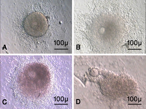
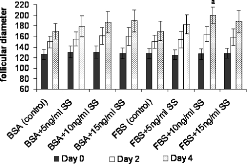
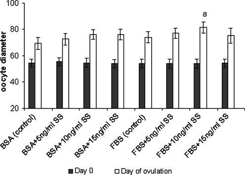
Methods: The isolated preantral follicles were cultured in TCM 199 medium supplemented with different concentrations (0, 5, 10, 15 ng/ml) of SS and 3 mg/ml bovine serum albumin (BSA) or 5% Fetal Bovine Serum (FBS). The ovulation was induced by addition of 1.5 IU/ml human chorionic gonadotropin. The size and development of follicles and oocytes were assessed by calibrated eyepiece.
Results: The survival rates of follicles in FBS supplemented groups containing 5 and 10 ng/ml SS (88.23%, 90.83%) were higher than other groups (P < 0.05 and P < 0.001 respectively). The mean diameter of follicles (199.84 +/- 15.58 microm) and the percentage of MII oocyte (33.08%) were higher in FBS supplemented group containing 10 ng/ml SS (P < 0.001).
Conclusion: The sodium selenite and FBS improve the in vitro growth and maturation of mouse preantral follicles.
Figures
Similar articles
-
In vitro maturation of bitch oocytes from advanced preantral follicles in synthetic oviduct fluid medium: serum is not essential.Theriogenology. 2002 Dec;58(9):1689-703. doi: 10.1016/s0093-691x(02)01080-4. Theriogenology. 2002. PMID: 12472139
-
Effect of activin A and insulin-like growth factor-I on in vitro development of preantral follicles isolated from cryopreserved ovarian tissues in the mouse.Cryobiology. 2008 Dec;57(3):209-15. doi: 10.1016/j.cryobiol.2008.08.004. Epub 2008 Aug 26. Cryobiology. 2008. PMID: 18789915
-
Development of morulae from the oocytes of cultured sheep preantral follicles.Theriogenology. 2010 Sep 15;74(5):884-94. doi: 10.1016/j.theriogenology.2010.04.013. Epub 2010 Jul 7. Theriogenology. 2010. PMID: 20615540
-
In vitro survival, growth, and maturation of sheep oocytes from secondary follicles cultured in serum-free conditions: impact of a constant or a sequential medium containing recombinant human FSH.Domest Anim Endocrinol. 2019 Apr;67:71-79. doi: 10.1016/j.domaniend.2018.12.003. Epub 2018 Dec 18. Domest Anim Endocrinol. 2019. PMID: 30765293
-
The base medium affects ultrastructure and survival of bovine preantral follicles cultured in vitro.Theriogenology. 2016 Apr 1;85(6):1019-29. doi: 10.1016/j.theriogenology.2015.11.007. Epub 2015 Nov 19. Theriogenology. 2016. PMID: 26711701 Review.
Cited by
-
Effects of Matricaria chamomilla Extract on Growth and Maturation of Isolated Mouse Ovarian Follicles in a Three-dimensional Culture System.Chin Med J (Engl). 2018 Jan 20;131(2):218-225. doi: 10.4103/0366-6999.222324. Chin Med J (Engl). 2018. PMID: 29336372 Free PMC article.
-
The effects of activated omental extract on nuclear and cytoplasmic in vitro maturation of rat oocytes.Iran J Basic Med Sci. 2017 Dec;20(12):1345-1353. doi: 10.22038/IJBMS.2017.9622. Iran J Basic Med Sci. 2017. PMID: 29238470 Free PMC article.
-
Sodium selenite improves folliculogenesis in radiation-induced ovarian failure: a mechanistic approach.PLoS One. 2012;7(12):e50928. doi: 10.1371/journal.pone.0050928. Epub 2012 Dec 6. PLoS One. 2012. PMID: 23236409 Free PMC article.
-
Fertility preservation among the cancer patients by ovarian tissue cryopreservation, transplantation, and follicular development.Iran J Cancer Prev. 2013 Summer;6(3):123-32. Iran J Cancer Prev. 2013. PMID: 25250122 Free PMC article. Review.
-
Ultrastructural and Morphalogical Changes of Mouse Ovarian Tissues Following Direct Cover Vitrification with Different Cryoprotectants.J Reprod Infertil. 2015 Jul-Sep;16(3):138-47. J Reprod Infertil. 2015. PMID: 26913232 Free PMC article.
References
-
- {'text': '', 'ref_index': 1, 'ids': [{'type': 'DOI', 'value': '10.1292/jvms.63.619', 'is_inner': False, 'url': 'https://doi.org/10.1292/jvms.63.619'}, {'type': 'PubMed', 'value': '11459007', 'is_inner': True, 'url': 'https://pubmed.ncbi.nlm.nih.gov/11459007/'}]}
- Bishonga C, Takahashi Y, Katagiri S, Nagano M, Ishikawa A. In vitro growth of mouse ovarian preantral follicles and the capacity of their oocytes to develop to blastocyst stage. J Vet Med Sci. 2001;63:619–624. doi:10.1292/jvms.63.619. - PubMed
-
- {'text': '', 'ref_index': 1, 'ids': [{'type': 'DOI', 'value': '10.1111/j.1447-0578.2007.00169.x', 'is_inner': False, 'url': 'https://doi.org/10.1111/j.1447-0578.2007.00169.x'}, {'type': 'PMC', 'value': 'PMC5906896', 'is_inner': False, 'url': 'https://pmc.ncbi.nlm.nih.gov/articles/PMC5906896/'}, {'type': 'PubMed', 'value': '29699267', 'is_inner': True, 'url': 'https://pubmed.ncbi.nlm.nih.gov/29699267/'}]}
- Hasegawa A. In vitro growth and maturation of mouse oocyte-granulosa cell complex from cryopreserved ovaries and achievement of pup birth. Reprod Med Biol. 2007;6:77–83. doi:10.1111/j.1447-0578.2007.00169.x. - PMC - PubMed
-
- Haidari K, Salehnia M, Valoujerdi MR. The effect of leukemia inhibitory factor and co-culture on the in vitro maturation and ultrastructure of vitrified and non-vitrified isolated mouse preantral follicles. Fertil Steril. 2008; In press. - PubMed
-
- {'text': '', 'ref_index': 1, 'ids': [{'type': 'PubMed', 'value': '9021369', 'is_inner': True, 'url': 'https://pubmed.ncbi.nlm.nih.gov/9021369/'}]}
- Cortvrindt R, Smitz J, Van Steirteghem AC. In vitro maturation, fertilization and embryo development of immature oocytes from early preantral follicles from prepuberal mice in a simplified culture system. Hum Reprod. 1996;11:2656–66. - PubMed
-
- {'text': '', 'ref_index': 1, 'ids': [{'type': 'DOI', 'value': '10.1095/biolreprod.105.042390', 'is_inner': False, 'url': 'https://doi.org/10.1095/biolreprod.105.042390'}, {'type': 'PMC', 'value': 'PMC2662519', 'is_inner': False, 'url': 'https://pmc.ncbi.nlm.nih.gov/articles/PMC2662519/'}, {'type': 'PubMed', 'value': '15987824', 'is_inner': True, 'url': 'https://pubmed.ncbi.nlm.nih.gov/15987824/'}]}
- Kreeger PK, Fernandes NN, Woodruff TK, Shea LD. Regulation of mouse follicle development by follicle-stimulating hormone in a three-dimensional in vitro culture system is dependent on follicle stage and dose. Biol Reprod. 2005;73:942–50. doi:10.1095/biolreprod.105.042390. - PMC - PubMed
Publication types
MeSH terms
Substances
LinkOut - more resources
Full Text Sources
