Comparative Study
. 2009 Mar;23(3):309-17.
doi: 10.1016/j.bbi.2008.09.002. Epub 2008 Sep 12.

Peripheral lipopolysaccharide (LPS) challenge promotes microglial hyperactivity in aged mice that is associated with exaggerated induction of both pro-inflammatory IL-1beta and anti-inflammatory IL-10 cytokines

Affiliations
Comparative Study

Peripheral lipopolysaccharide (LPS) challenge promotes microglial hyperactivity in aged mice that is associated with exaggerated induction of both pro-inflammatory IL-1beta and anti-inflammatory IL-10 cytokines

Christopher J Henry et al. Brain Behav Immun. 2009 Mar.

Abstract

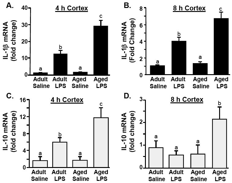
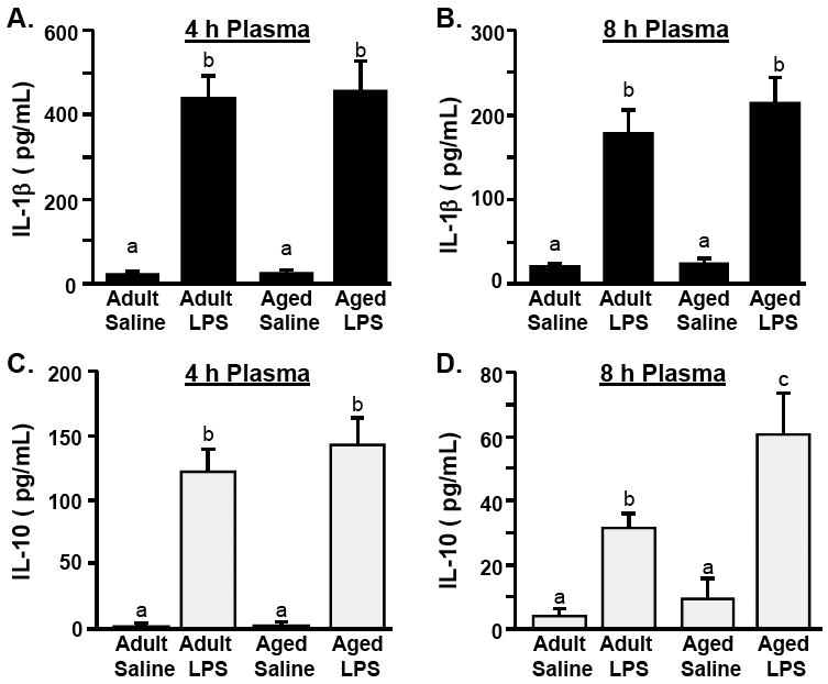
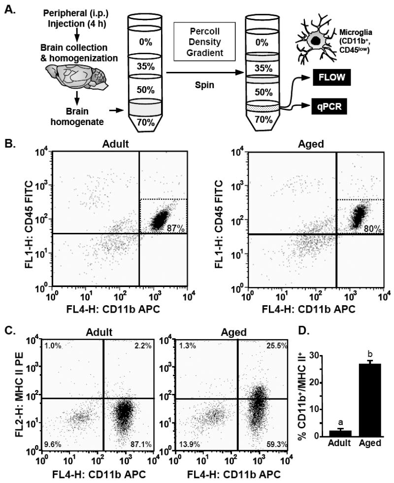
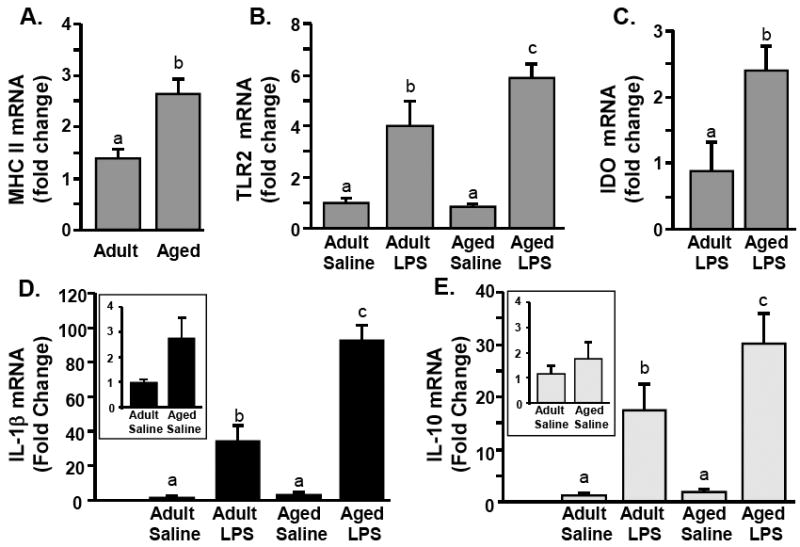
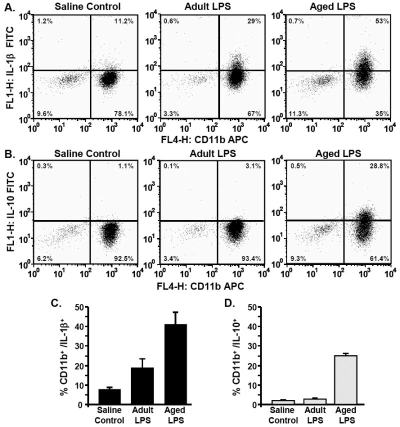
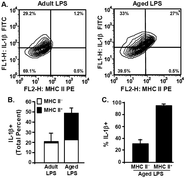
In the elderly, systemic infection is associated with an increased frequency of behavioral and cognitive complications. We have reported that peripheral stimulation of the innate immune system with lipopolysaccharide (LPS) causes an exaggerated neuroinflammatory response and prolonged sickness/depressive-like behaviors in aged BALB/c mice. Therefore, the purpose of this study was to determine the degree to which LPS-induced neuroinflammation was associated with microglia-specific induction of neuroinflammatory mediators. Here, we show that peripheral LPS challenge caused a hyperactive microglial response in the aged brain associated with higher induction of inflammatory IL-1beta and anti-inflammatory IL-10. LPS injection caused a marked induction of mRNA expression of both IL-1beta and IL-10 in the cortex of aged mice compared to adults. In the next set of studies, microglia (CD11b(+)/CD45(low)) were isolated from the brain of adult and aged mice following experimental treatments. An age-dependent increase in major histocompatibility complex (MHC) class II mRNA and protein expression was detected in microglia. Moreover, peripheral LPS injection caused a more pronounced increase in IL-1beta, IL-10, Toll-like receptor (TLR)-2, and indoleamine 2,3-dioxygenase (IDO) mRNA levels in microglia isolated from aged mice than adults. Intracellular cytokine protein detection confirmed that peripheral LPS caused the highest increase in IL-1beta and IL-10 levels in microglia of aged mice. Finally, the most prominent induction of IL-1beta was detected in MHC II(+) microglia from aged mice. Taken together, these findings provide novel evidence that age-associated priming of microglia plays a central role in exaggerated neuroinflammation induced by activation of the peripheral innate immune system.


Figures

Fig 1
Fig 1. Peripheral LPS injection causes exaggerated mRNA expression of IL-1β and IL-10 in the cortex of aged mice compared to adults
Adult (3-6 m) and aged (18-20 m) mice were challenged with saline or LPS i.p. and IL-1β mRNA levels were determined from the cortex collected A) 4 or B) 8 h later. From the same cortex samples, IL-10 mRNA levels were determined C) 4 or D) 8 h post experimental treatments. Bars represent the mean ± SEM (n=8 for 4 h, n=5 for 8 h). Means with different letters (a, b, or c) are significantly different (P<0.05) from each other.
Fig.2
Fig.2. LPS injection increases IL-1β and IL-10 and in the plasma independent of age
Adult (3-6 m) and aged (18-20 m) mice were challenged with saline or LPS i.p. and IL-1β protein levels were determined from the plasma collected A) 4 h or B) 8 h later. From the same plasma samples, IL-10 protein levels were determined C) 4 or D) 8 h post experimental treatments. Bars represent the mean ± SEM (n=8 for 4 h, n=5 for 8 h). Means with different letters (a, b, or c) are significantly different (P<0.05) from each other.
Fig.3
Fig.3. Increased MHC II surface expression on microglia isolated from the brain of aged mice
A) Illustration of the experimental protocol used to isolated microglia from adult (3-6 m) and aged (18-20 m) mice by Percoll density gradient. B) Representative bivariate dot plots of Percoll isolated cells stained with anti-CD11b-APC and anti-CD45-FITC. Microglia were identified by CD11b+/CD45low staining. C) Representative bivariate dot plots of Percoll isolated cells stained with anti-CD11b-APC and anti-MHC-II-PE. D) Average percentage of cells that were CD11b+/MHC II+. Bars represent the mean ± SEM (n=8). Means with different letters (a or b) are significantly different (P<0.05) from each other.
Fig.4
Fig.4. Peripheral LPS injection causes exaggerated mRNA levels of inflammatory mediators in microglia of aged mice compared to adults
Adult (3-6 m) and aged (18-20 m) mice were challenged with saline or LPS i.p. and A) MHC II, B) TLR2, C) IDO, D) IL-1β and E) IL-10 mRNA levels were determined from enriched microglia isolated 4 h later. Bars represent the mean ± SEM (n=8). Means with different letters (a, b, or c) are significantly different (P<0.05) from each other.
Fig.5
Fig.5. Higher percentage of IL-1β+ and IL-10 positive microglia in aged mice compared to adults following peripheral LPS injection
Adult (3-6 m) and aged (18-20 m) mice were challenged with saline or LPS i.p. and microglia were isolated 4 h later by Percoll density gradient. Cells were subjected to the BD Cytofix/Cytoperm Plus fixation/permeabilization protocol as described in the Methods. Representative bivariate dot plots of Percoll isolated cells stained with A) anti-CD11b-APC and anti-IL-1β-FITC or B) anti-CD11b-APC and anti-IL-10-FITC. Average percentage of cells that were C) CD11b+/IL-1β+ and D) CD11b+/IL-10+. Bars represent the mean ± SEM (n=3-6).
Fig.6
Fig.6. MHC II positive microglia from aged mice have a robust IL-1β induction following peripheral LPS injection
A subset of Percoll isolated microglia (Fig.5) were also stained with anti-MHC-II-PE. Cells were gated by CD11b+ staining. A) Representative bivariate contour plots of CD11b+ cells that were stained with anti-IL-1β-FITC and anti-MHC-II-PE. B) Average total percent of microglia that were IL-1β+ following peripheral LPS injection, differentiated based on whether they were MHC IIneg (white bars) or MHC II+ (black bars). Bars represent the mean ± SEM (n=3). C) Average percent of MHC IIneg and MHC II+ microglia that were also IL-1β+. Bars represent the mean ± SEM (n=3).

References

    1. Abraham J, Jang S, Godbout JP, Chen J, Kelley KW, Dantzer R, Johnson RW. Aging sensitizes mice to behavioral deficits induced by central HIV-1 gp120. Neurobiol Aging. 2008;29(4):614–21. - PMC - PubMed
    1. Allan SM, Tyrrell PJ, Rothwell NJ. Interleukin-1 and neuronal injury. Nat Rev Immunol. 2005;5(8):629–40. - PubMed
    1. Amaral ME, Barbuio R, Milanski M, Romanatto T, Barbosa HC, Nadruz W, Bertolo MB, Boschero AC, Saad MJ, Franchini KG, Velloso LA. Tumor necrosis factor-alpha activates signal transduction in hypothalamus and modulates the expression of pro-inflammatory proteins and orexigenic/anorexigenic neurotransmitters. J Neurochem. 2006;98(1):203–12. - PubMed
    1. Barrientos RM, Higgins EA, Biedenkapp JC, Sprunger DB, Wright-Hardesty KJ, Watkins LR, Rudy JW, Maier SF. Peripheral infection and aging interact to impair hippocampal memory consolidation. Neurobiol Aging. 2006;27(5):723–32. - PubMed
    1. Basak C, Pathak SK, Bhattacharyya A, Mandal D, Pathak S, Kundu M. NF-kappaB- and C/EBPbeta-driven interleukin-1beta gene expression and PAK1-mediated caspase-1 activation play essential roles in interleukin-1beta release from Helicobacter pylori lipopolysaccharide-stimulated macrophages. J Biol Chem. 2005;280(6):4279–88. - PubMed

Publication types

MeSH terms