

Subregional hippocampal atrophy predicts Alzheimer's dementia in the cognitively normal
- PMID: 18814937
- PMCID: PMC2873083
- DOI: 10.1016/j.neurobiolaging.2008.08.008
Subregional hippocampal atrophy predicts Alzheimer's dementia in the cognitively normal
Abstract
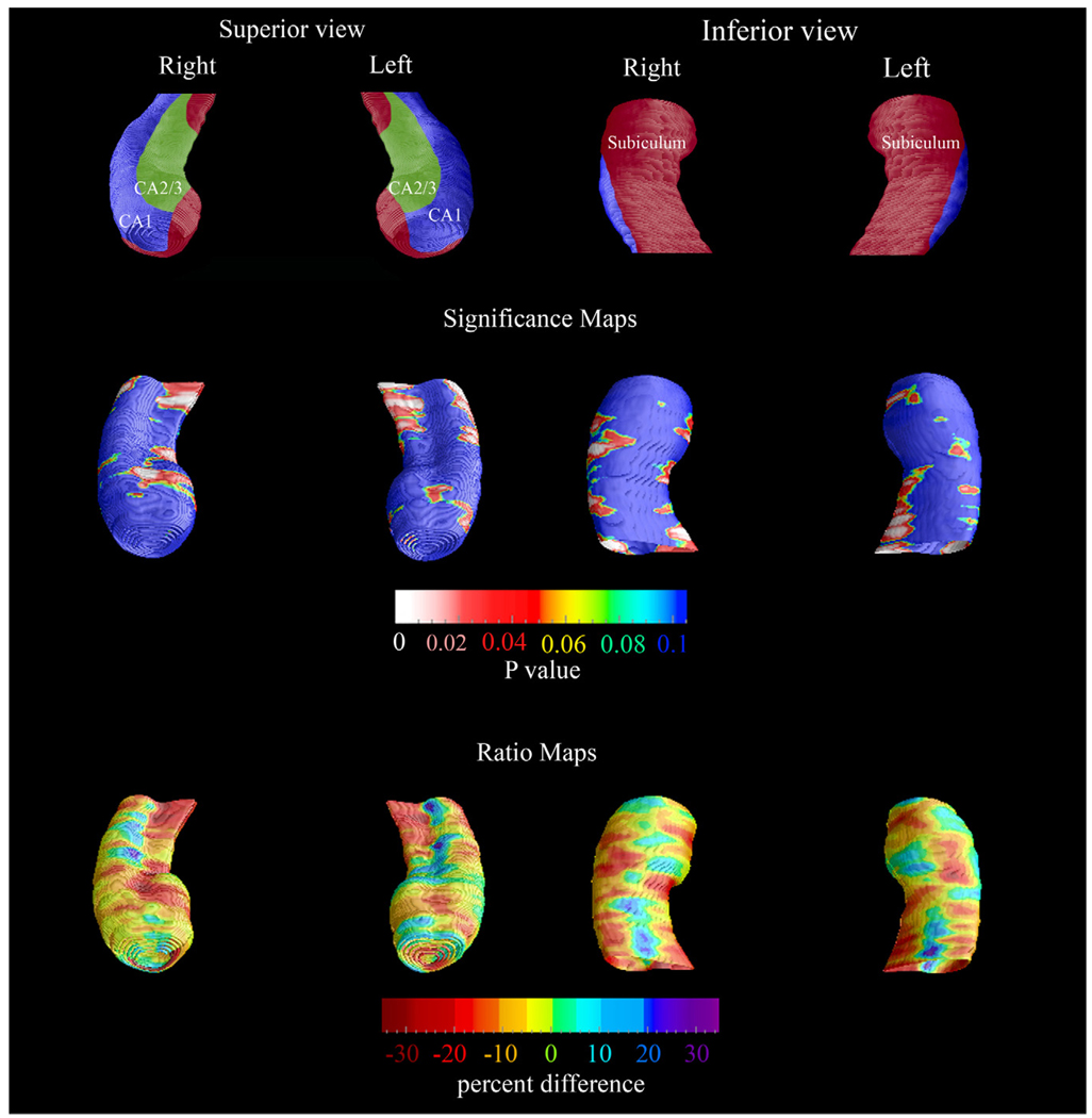
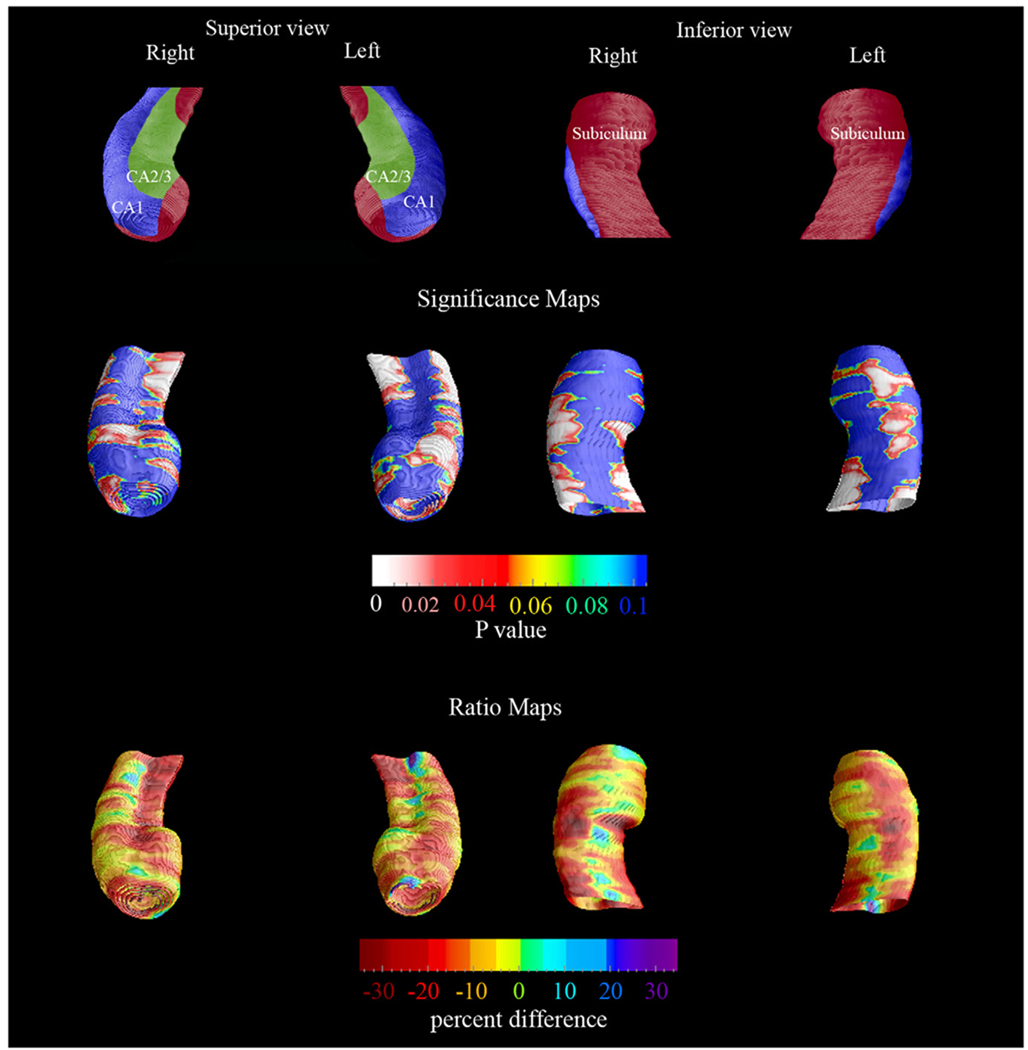
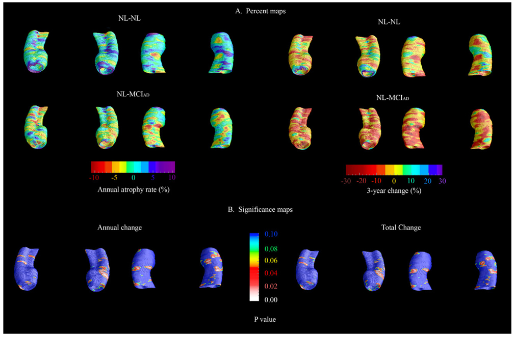
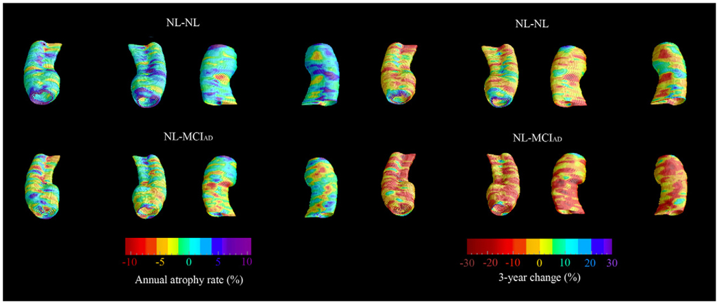
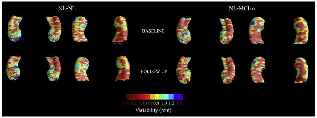
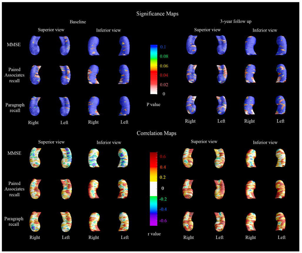
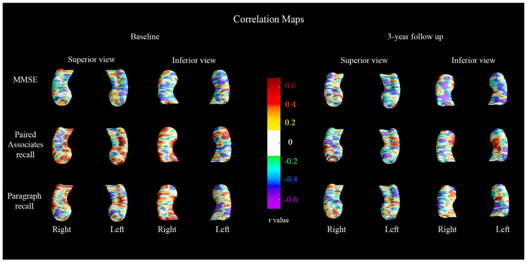
Atrophic changes of the hippocampus are typically regarded as an early sign of Alzheimer's dementia (AD). Using the radial distance atrophy mapping approach, we compared the longitudinal MRI data of 10 cognitively normal elderly subjects who remained normal at 3-year and 6-year follow-up (NL-NL) and 7 cognitively normal elderly subjects who were diagnosed with mild cognitive impairment (MCI) 2.8 (range 2.0-3.9) and with AD 6.8 years (range 6.1-8.2) after baseline (NL-MCI(AD)). 3D statistical maps revealed greater hippocampal atrophy in the NL-MCI(AD) relative to the NL-NL group at baseline (left p=0.05; right p=0.06) corresponding to 10-15% CA1, and 10-25% subicular atrophy, and bilateral differences at 3-year follow-up (left p=0.001, right p<0.02) corresponding to 10-30% subicular, 10-20% CA1, and 10-20% newly developed CA2-3 atrophy. This preliminary study suggests that excess CA1 and subicular atrophy is present in cognitively normal individuals predestined to decline to amnestic MCI, while progressive involvement of the CA1 and subiculum, and atrophy spreading to the CA2-3 subfield in amnestic MCI, suggests future diagnosis of AD.
Copyright 2008 Elsevier Inc. All rights reserved.
Conflict of interest statement
Dr. Thompson, Ms. Green and Ms. Mistur have no conflicts of interest. Ms. Hwang and Mr. Ramirez have nothing to disclose.
Figures

References
-
- Apostolova LG, Dinov ID, Dutton RA, Hayashi KM, Toga AW, Cummings JL, Thompson PM. 3D comparison of hippocampal atrophy in amnestic mild cognitive impairment and Alzheimer’s disease. Brain. 2006a;129(Pt 11):2867–2873. - PubMed
-
- Apostolova LG, Dutton RA, Dinov ID, Hayashi KM, Toga AW, Cummings JL, Thompson PM. Conversion of mild cognitive impairment to Alzheimer disease predicted by hippocampal atrophy maps. Arch. Neurol. 2006b;63(5):693–699. - PubMed
-
- Becker JT, Davis SW, Hayashi KM, Meltzer CC, Toga AW, Lopez OL, Thompson PM. Three-dimensional patterns of hippocampal atrophy in mild cognitive impairment. Arch. Neurol. 2006;63(1):97–101. - PubMed
-
- Bobinski M, de Leon MJ, Tarnawski M, Wegiel J, Reisberg B, Miller DC, Wisniewski HM. Neuronal and volume loss in CA1 of the hippocampal formation uniquely predicts duration and severity of Alzheimer disease. Brain Res. 1998;805(1–2):267–269. - PubMed
Publication types
MeSH terms
Grants and funding
- R01 AG022374/AG/NIA NIH HHS/United States
- P50 AG16570/AG/NIA NIH HHS/United States
- R01 AG013616/AG/NIA NIH HHS/United States
- P30 AG008051/AG/NIA NIH HHS/United States
- R01 AG012101/AG/NIA NIH HHS/United States
- K23 AG026803/AG/NIA NIH HHS/United States
- U54 RR021813/RR/NCRR NIH HHS/United States
- LM05639/LM/NLM NIH HHS/United States
- AG13616/AG/NIA NIH HHS/United States
- R01 MH071940/MH/NIMH NIH HHS/United States
- AG08051/AG/NIA NIH HHS/United States
- AG12101/AG/NIA NIH HHS/United States
- R21 RR019771/RR/NCRR NIH HHS/United States
- R01 LM005639/LM/NLM NIH HHS/United States
- RR019771/RR/NCRR NIH HHS/United States
- P50 AG016570/AG/NIA NIH HHS/United States
- AG022374/AG/NIA NIH HHS/United States
- P41 RR013642/RR/NCRR NIH HHS/United States
LinkOut - more resources
Full Text Sources
Other Literature Sources
Medical
Miscellaneous
