. 2008 Sep 24;28(39):9692-701.
doi: 10.1523/JNEUROSCI.1551-08.2008.

Numbers, densities, and colocalization of AMPA- and NMDA-type glutamate receptors at individual synapses in the superficial spinal dorsal horn of rats

Affiliations

Numbers, densities, and colocalization of AMPA- and NMDA-type glutamate receptors at individual synapses in the superficial spinal dorsal horn of rats

Miklós Antal et al. J Neurosci. .

Abstract

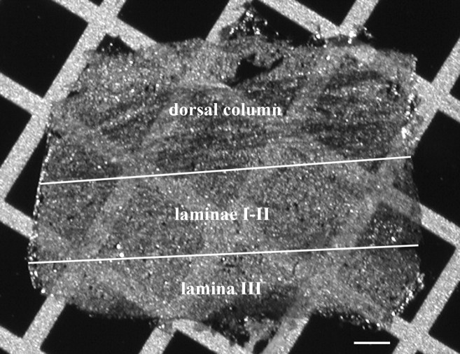
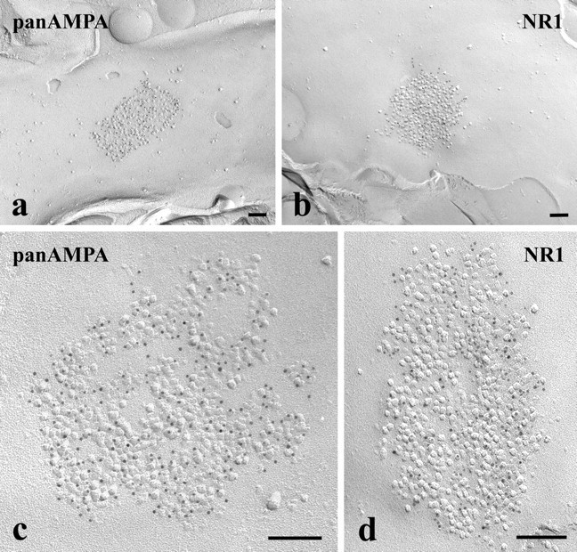
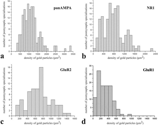
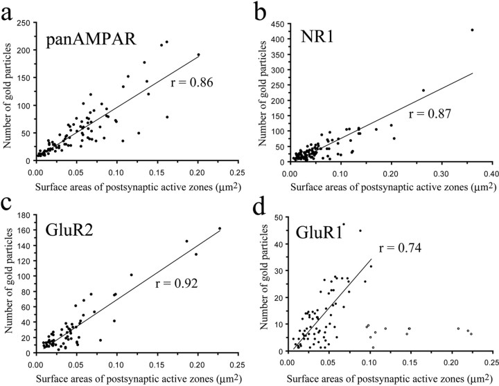
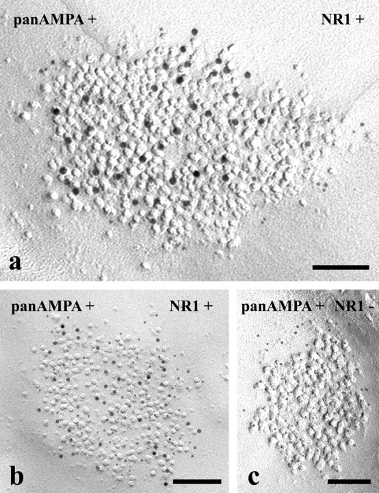
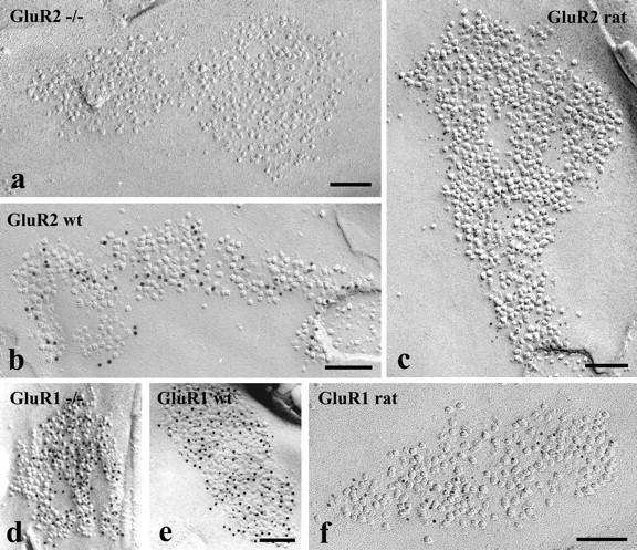
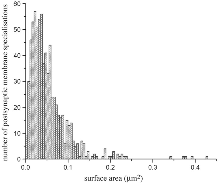
Ionotropic glutamate receptors play important roles in spinal processing of nociceptive sensory signals and induction of central sensitization in chronic pain. Here we applied highly sensitive freeze-fracture replica labeling to laminae I-II of the spinal dorsal horn of rats and investigated the numbers, densities, and colocalization of AMPA- and NMDA-type glutamate receptors at individual postsynaptic membrane specializations with a high resolution. All glutamatergic postsynaptic membranes in laminae I-II expressed AMPA receptors, and most of them (96%) were also immunoreactive for the NR1 subunit of NMDA receptors. The numbers of gold particles for AMPA and NMDA receptors at individual postsynaptic membranes showed a linear correlation with the size of postsynaptic membrane specializations and varied in the range of 8-214 and 5-232 with median values of 37 and 28, whereas their densities varied in the range of 325-3365/microm(2) and 102-2263/microm(2) with median values of 1115/microm(2) and 777/microm(2), respectively. Virtually all (99%) glutamatergic postsynaptic membranes expressed GluR2, and most of them (87%) were also immunoreactive for GluR1. The numbers of gold particles for pan-AMPA, NR1, and GluR2 subunits showed a linear correlation with the size of postsynaptic surface areas. Concerning GluR1, there may be two populations of synapses with high and low GluR1 densities. In synapses larger than 0.1 microm(2), GluR1 subunits were recovered in very low numbers. Differential expression of GluR1 and GluR2 subunits suggests regulation of AMPA receptor subunit composition by presynaptic mechanism.


Figures

Figure 1.
Figure 1.
Photomicrograph of an SDS-FRL replica on a 100-mesh copper grid. The replica was prepared from a paramedian–sagittal section of the dorsal aspect of the rat lumbar spinal cord. White lines indicate the presumed outer and inner borders of laminae I–II. Scale bar, 100 μm.
Figure 2.
Figure 2.
a–d, Electron micrographs of SDS-FRL replicas immunolabeled with an antibody that recognizes all subunits of AMPA-type glutamate receptors (pan-AMPA) (a, c) or with an antibody raised against the NR1 subunit of NMDA-type glutamate receptors (b, d). The micrographs illustrate postsynaptic membrane specializations (IMP clusters) that show strong immunoreactivity for pan-AMPA (a, c) or NR1 (b, d) in the superficial spinal dorsal horn of rats. All subunits are labeled with 5 nm gold particles. Scale bars, 0.1 μm.
Figure 3.
Figure 3.
a–d, Histograms showing the distribution of densities of gold particles that label various subunits of AMPA- (a, c, d) and NMDA- (b) type glutamate receptors in SDS-FRL replicas of individual postsynaptic membrane specializations in the superficial spinal dorsal horn of rats. Replicas from which data were obtained were immunolabeled with antibodies that recognize all subunits (pan-AMPA) (a) of AMPA receptors, NR1 subunits of NMDA receptors (b), and GluR2 (c) and GluR1 (d) subunits of AMPA receptors.
Figure 4.
Figure 4.
a–d, Scatter plots showing the correlation between surface areas of postsynaptic membrane specializations and numbers of gold particles labeling all subunits (pan-AMPA) (a) of AMPA receptors, NR1 subunit of NMDA receptors (b), and GluR2 (c) and GluR1 (d) subunits of AMPA receptors. Open circles in d label data from postsynaptic membrane specializations that are larger than 0.06 μm2 and present low numbers of gold particles. These synapses were excluded from the calculation of the value of linear correlation.
Figure 5.
Figure 5.
Electron micrographs of SDS-FRL replicas simultaneously double immunolabeled with antibodies raised against the NR1 subunit of NMDA-type glutamate receptors and an amino acid sequence that is common in all subunits of AMPA-type glutamate receptors (pan-AMPA). a–c, AMPA receptors are labeled with 10 nm gold particles, and NR1 subunits are marked with 5 nm gold particles in a, whereas 5 nm gold particles label AMPA receptors, and 10 nm gold particles mark NR1 subunits in b and c. Postsynaptic membrane specializations in the micrographs in a and b show immunoreactivity for both antibodies, whereas postsynaptic membrane specializations in the micrograph in c are positive for pan-AMPA but negative for NR1. Scale bars, 0.1 μm.
Figure 6.
Figure 6.
Electron micrographs of SDS-FRL replicas immunolabeled with antibodies that recognize GluR2 or GluR1 subunits. a–f, The micrographs illustrate postsynaptic membrane specializations (IMP clusters) in the superficial spinal dorsal horn of GluR2 (a) and GluR1 (d) knock-out mice and wild-type (wt) mice (b, e) and rats (c, f). SDS-FRL replicas from GluR2 knock-out mice (a) and wild-type mice (b) and rats (c) were labeled for GluR2. The postsynaptic membrane specializations are free of labeling in the GluR2 knock-out animal (a), and show strong immunoreactivity for GluR2 in the wild-type mice (b) and rats (c). SDS-FRL replicas from GluR1 knock-out (d) and wild-type (e) mice were double labeled for GluR1 and GluR2, whereas replicas from a rat were labeled only for GluR1 (f). The postsynaptic membrane specializations from GluR1 knock-out animals are positively labeled for GluR2 but negative for GluR1 (d). The postsynaptic membrane specialization from wild-type mice are positive for both GluR1 and GluR2 (e), whereas postsynaptic membrane specialization from a rat show strong immunoreactivity for GluR1 (f). GluR2 subunits were labeled with 10 nm (b, d, e) and 5 nm (c), whereas GluR1 subunits were labeled with 5 nm (d, e) and 10 nm (f) gold particles. Scale bars, 0.1 μm.
Figure 7.
Figure 7.
Histogram showing the distribution of surface areas of complete postsynaptic membrane specializations expressing AMPA- and/or NMDA-type glutamate receptors in the superficial spinal dorsal horn of rats.

References

    1. Albuquerque C, Lee CJ, Jackson AC, MacDermott AB. Subpopulations of GABAergic and non-GABAergic rat dorsal horn neurons express Ca2+-permeable AMPA receptors. Eur J Neurosci. 1999;11:2758–2766. - PubMed
    1. Antal M, Kraftsik R, Székely G, van der Loos H. Synapses on motoneuron dendrites in the brachial section of the frog spinal cord: a computer-aided electron microscopic study of cobalt-filled cells. J Neurocytol. 1992;21:34–49. - PubMed
    1. Baba H, Doubell TP, Moore KA, Woolf CJ. Silent NMDA receptor-mediated synapses are developmentally regulated in the dorsal horn of the rat spinal cord. J Neurophysiol. 2000;83:955–962. - PubMed
    1. Bardoni R, Magherini PC, MacDermott AB. NMDA EPSCs at glutamatergic synapses in the spinal cord dorsal horn of the postnatal rat. J Neurosci. 1998;18:6558–6567. - PMC - PubMed
    1. Baude A, Nusser Z, Molnár E, McIlhinney RA, Somogyi P. High-resolution immunogold localization of AMPA type glutamate receptor subunits at synaptic and non-synaptic sites in rat hippocampus. Neuroscience. 1995;69:1031–1055. - PubMed

Publication types

Substances