. 2008 Dec;82(23):11651-68.
doi: 10.1128/JVI.01762-08. Epub 2008 Sep 24.

Profiling the specificity of neutralizing antibodies in a large panel of plasmas from patients chronically infected with human immunodeficiency virus type 1 subtypes B and C

Affiliations

Profiling the specificity of neutralizing antibodies in a large panel of plasmas from patients chronically infected with human immunodeficiency virus type 1 subtypes B and C

James M Binley et al. J Virol. 2008 Dec.

Abstract

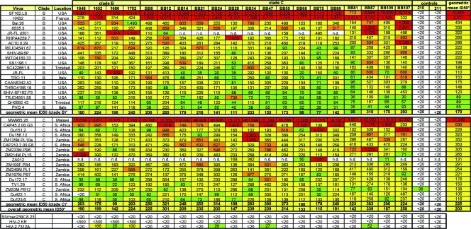
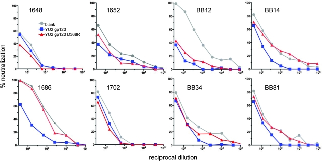
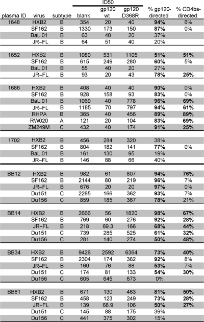
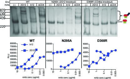
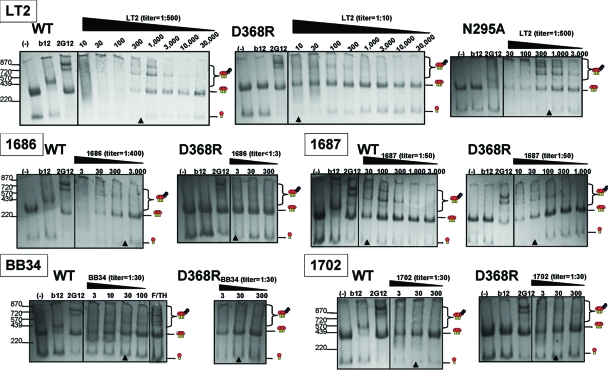
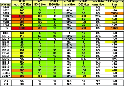
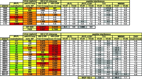
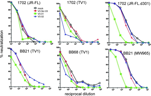
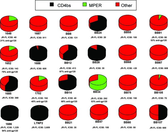
Identifying the viral epitopes targeted by broad neutralizing antibodies (NAbs) that sometimes develop in human immunodeficiency virus type 1 (HIV-1)-infected subjects should assist in the design of vaccines to elicit similar responses. Here, we investigated the activities of a panel of 24 broadly neutralizing plasmas from subtype B- and C-infected donors using a series of complementary mapping methods, focusing mostly on JR-FL as a prototype subtype B primary isolate. Adsorption with gp120 immobilized on beads revealed that an often large but variable fraction of plasma neutralization was directed to gp120 and that in some cases, neutralization was largely mediated by CD4 binding site (CD4bs) Abs. The results of a native polyacrylamide gel electrophoresis assay using JR-FL trimers further suggested that half of the subtype B and a smaller fraction of subtype C plasmas contained a significant proportion of NAbs directed to the CD4bs. Anti-gp41 neutralizing activity was detected in several plasmas of both subtypes, but in all but one case, constituted only a minor fraction of the overall neutralization activity. Assessment of the activities of the subtype B plasmas against chimeric HIV-2 viruses bearing various fragments of the membrane proximal external region (MPER) of HIV-1 gp41 revealed mixed patterns, implying that MPER neutralization was not dominated by any single specificity akin to known MPER-specific monoclonal Abs. V3 and 2G12-like NAbs appeared to make little or no contribution to JR-FL neutralization titers. Overall, we observed significant titers of anti-CD4bs NAbs in several plasmas, but approximately two-thirds of the neutralizing activity remained undefined, suggesting the existence of NAbs with specificities unlike any characterized to date.


Figures

FIG. 1.
FIG. 1.
Breadth and potency of neutralization by a panel of subtype B and C plasmas. Four of the 8 subtype B and all 16 subtype C plasmas, organized horizontally, were evaluated in the TZM-bl neutralization assay, with the exception of JR-FL neutralization that was determined in the CF2 assay. A panel of subtype B and C viruses are organized in the top and bottom halves of this figure, respectively, and control HIV-2 and SIV viruses are shown at the bottom. Subtype B and C viruses are arranged with those for the most-sensitive tier 1 isolates at the top and the most-resistant tier 2 isolates at the bottom, as shown by calculations of the geometric mean titers in the rightmost column. The country of origin of each Env, where known, is indicated in the third column. ID50 titers are color coded as follows: white boxes, titer <20; green boxes, titer <100; yellow boxes, titer <500; orange boxes, titer <1,000; and red boxes, titer >1,000. *, calculations of geometric mean titers took into account values against all isolates except the overtly sensitive JR-FL.d301, SF162.LS, HXB2, and MW965.26 viruses. n.d., not done.
FIG. 2.
FIG. 2.
Neutralization analysis of plasmas fractionated on gp120 beads. Plasmas were fractionated on YU2 WT gp120, D368R gp120, or blank (control) beads. JR-FL neutralization assays were performed on unbound fractions using the TZM-bl assay.
FIG. 3.
FIG. 3.
Summary of plasma fractionation on gp120 beads. Plasmas at a 1:20 dilution were adsorbed to blank or gp120 beads and then tested in TZM-bl neutralization assays, causing a further twofold dilution, giving a final starting dilution of 1:40. Reciprocal neutralization ID50 titers are shown. In cases where no neutralization was observed at 1:40, a value of 20 is given. Fourth column, plasmas adsorbed on untreated tosyl-activated Dynabeads; fifth column, plasmas adsorbed with WT YU2 gp120; sixth column, plasmas adsorbed with D368R YU2 gp120 (CD4bs knockout mutation); seventh column, percent reduction in total neutralizing ID50 by WT gp120 adsorption relative to that of blank beads. In cases where the WT titer was higher than that of the blank, a value of 0% was assigned; in cases where gp120-directed activity was >50% of the total neutralizing titer, the contribution of CD4bs activity was calculated. Eighth column, CD4bs-directed activity expressed as the percentage of the total neutralization titer. Values in bold are considered significant; i.e., cases where gp120 adsorbs >50% of total neutralizing activity and >25% of the activity is directed to the CD4bs. The calculations underlying these percentages are described in Materials and Methods.
FIG. 4.
FIG. 4.
Evaluation of WT and mutant JR-FL Envs in BN-PAGE and infectivity assays. Upper panel, WT, N295A (2G12 knockout mutant), and D368R (CD4bs knockout mutant) particles were incubated alone (−) or with one of the MAbs 2G12, IgG1b12, and sCD4 at 30 μg/ml. Trimers were then separated in BN-PAGE. Molecular mass markers for ferritin (220 and 439 kDa) and the estimated sizes of 2G12-liganded trimers (∼570, ∼720, and ∼870 kDa) are indicated. Lower panel, WT and mutant particle sensitivities to neutralization by MAbs IgG1b12 and 2G12 were evaluated in neutralization assays with CF2 target cells. RLU, relative light units.
FIG. 5.
FIG. 5.
Evaluation of plasma binding to WT and mutant trimers by BN-PAGE. Plasmas were titrated against WT, D368R, or N295A trimers in BN-PAGE as indicated. Cartoons depict gp120/gp41 trimers, trimer-IgG complexes, and monomers. Markers show the locations of ferritin markers of 220 and 439 kDa and trimers complexed with one, two, or three molecules of IgG 2G12 (molecular masses of ∼570, ∼720, and ∼870 kDa). Arrows indicate the dilution corresponding to the ID50 titer, inferred from at least three duplicate titrations. The unbound fraction of plasma BB34 absorbed against WT gp120 beads was also evaluated for plasma BB34, identified as “F/TH,” flowthrough. (−), blank beads.
FIG. 6.
FIG. 6.
Summary of results of mapping by native PAGE. Color coding is exactly as described in the Fig. 1 legend. ID50 neutralization (neut.) titers against JR-FL measured in the CF2 assay are indicated in the second column. ID50 titers of binding to WT, D368R, and N295A mutant titers are shown in the third, fourth, and sixth columns, respectively. The percentage of D368R (CD4bs)- or N295A (2G12-like)-sensitive activity was calculated with reference to the total activity against WT trimers (fifth and seventh columns). Gray-shaded cells in the fifth and seventh columns are considered significant, i.e., the titers differ by more than twofold, leading to a 50% or greater contribution to neutralization. ID50 competition titers for virus capture by the 2G12 MAb are shown in the eighth column. n.d., not done; N/A, not applicable.
FIG. 7.
FIG. 7.
Summary of MPER-neutralizing activity. MPER ID50 titers were measured in post-CD4/CCR5 and HIV-2 MPER graft neutralization assays. Post-CD4/CCR5 activity (third column) is expressed as a percentage, given to the nearest integer (fourth column), of that in the standard format (second column), both assayed on CF2 cells. Titers against the YU2 C1 MPER/HIV-2 chimera virus (fifth column) have been adjusted to subtract the HIV-2 7312A background titers (Fig. 1), so that all the activity is likely to be directed against the MPER. The IC50s of MPER MAbs 2F5, 4E10, and Z13e1 were also determined in each assay, expressed in μg/ml. Boxes have color coding similar to that in Fig. 1; gray-shaded boxes in the fourth column indicate samples with significant MPER activity. N/A, not applicable.
FIG. 8.
FIG. 8.
Summary of V3 activity. V3 activity was measured by (i) competition of virus capture by a V3 MAb 39F (third column), (ii) neutralization of the HIV-2 KR virus engrafted with either the YU2 or a consensus subtype C V3 loop (fourth and fifth columns), and (iii) by peptide interference assays (6th to 14th columns). Virus capture and chimera neutralization data are given as ID50 titers. Peptide interference was recorded as the severalfold change in ID50 titer with reference to the titer of controls to which no peptide was added. Virus capture and chimera neutralization assay data are color coded as described in the Fig. 1 legend. Boxes depicting the results of peptide interference experiments in which significant competition was observed (more-than-twofold effect) are shaded gray; results indicating no significant competition (less-than-twofold effect) are unshaded. Controls included V3 MAbs 447-52D, F425, and guinea pig serum WGP 102-4 (66). n.d., not done.
FIG. 9.
FIG. 9.
Roles of the V3 N and C termini in subtype B and C neutralization. Viruses and plasmas were incubated in the presence or absence of 25 μg/ml of the interfering peptide indicated, and neutralization was subsequently measured using the TZM-bl assay. In each experiment, V3 peptides (depicted in Fig. S6 in the supplemental material) were matched to the sequence of the virus used in neutralization assays. The “V3 Scr.03” peptide was included as an assay control. Its sequence, NKGTHNIPTARNIYGFPTSRRGT, is based on jumbled V3 residues.
FIG. 10.
FIG. 10.
Summary of estimated contributions of plasma specificities involved in JR-FL neutralization. CD4bs activities are derived from the trimer shift data shown in Fig. 6, and MPER activities are derived from the post-CD4/CCR5 activities shown in Fig. 7. Any remaining activities unaccounted for by mapping are depicted as “other.” Due to uncertainty regarding activity that is <50% of the total in native PAGE and peptide interference assays (Fig. 8), we only include data showing a >50% contribution to total neutralization, thus eliminating possible minor contributions by V3- and 2G12-like NAbs in these pie charts. The percentages of gp120-specific neutralization are derived from the data shown in Fig. 3.

References

    1. Abrahamyan, L. G., R. M. Markosyan, J. P. Moore, F. S. Cohen, and G. B. Melikyan. 2003. Human immunodeficiency virus type 1 Env with an intersubunit disulfide bond engages coreceptors but requires bond reduction after engagement to induce fusion. J. Virol. 775829-5836. - PMC - PubMed
    1. Bibollet-Ruche, F., L. Hui, J. M. Decker, P. A. Geopfert, B. H. Hahn, E. Delaporte, M. Peeters, S. Allen, E. Hunter, J. Robinson, P. D. Kwong, and G. M. Shaw. 2006. Detection of novel neutralizing antibody responses to the membrane proximal external region (MPER) or gp41 following infection by HIV-1 subtypes A, B, C, D, F, G, H, CRF01, CRF02, or CRF11, abstr. 110, p. 196. Keystone Symposium X6, HIV Vaccines, Keystone, CO, 27 March to 2 April.
    1. Binley, J. M., C. S. Cayanan, C. Wiley, N. Schulke, W. C. Olson, and D. R. Burton. 2003. Redox-triggered infection by disulfide-shackled human immunodeficiency virus type 1 pseudovirions. J. Virol. 775678-5684. - PMC - PubMed
    1. Binley, J. M., P. J. Klasse, Y. Cao, I. Jones, M. Markowitz, D. D. Ho, and J. P. Moore. 1997. Differential regulation of the antibody responses to Gag and Env proteins of human immunodeficiency virus type 1. J. Virol. 712799-2809. - PMC - PubMed
    1. Binley, J. M., T. Wrin, B. Korber, M. B. Zwick, M. Wang, C. Chappey, G. Stiegler, R. Kunert, S. Zolla-Pazner, H. Katinger, C. J. Petropoulos, and D. R. Burton. 2004. Comprehensive cross-clade neutralization analysis of a panel of anti-human immunodeficiency virus type 1 monoclonal antibodies. J. Virol. 7813232-13252. - PMC - PubMed

Publication types

MeSH terms

Substances