. 2008 Dec;82(23):11869-79.
doi: 10.1128/JVI.01634-08. Epub 2008 Sep 24.

Mutational analysis of cis-acting RNA signals in segment 7 of influenza A virus

Affiliations

Mutational analysis of cis-acting RNA signals in segment 7 of influenza A virus

Edward C Hutchinson et al. J Virol. 2008 Dec.

Abstract

The genomic viral RNA (vRNA) segments of influenza A virus contain specific packaging signals at their termini that overlap the coding regions. To further characterize cis-acting signals in segment 7, we introduced synonymous mutations into the terminal coding regions. Mutation of codons that are normally highly conserved reduced virus growth in embryonated eggs and MDCK cells between 10- and 1,000-fold compared to that of the wild-type virus, whereas similar alterations to nonconserved codons had little effect. In all cases, the growth-impaired viruses showed defects in virion assembly and genome packaging. In eggs, nearly normal numbers of virus particles that in aggregate contained apparently equimolar quantities of the eight segments were formed, but with about fourfold less overall vRNA content than wild-type virions, suggesting that, on average, fewer than eight segments per particle were packaged. Concomitantly, the particle/PFU and segment/PFU ratios of the mutant viruses showed relative increases of up to 300-fold, with the behavior of the most defective viruses approaching that predicted for random segment packaging. Fluorescent staining of infected cells for the nucleoprotein and specific vRNAs confirmed that most mutant virus particles did not contain a full genome complement. The specific infectivity of the mutant viruses produced by MDCK cells was also reduced, but in this system, the mutations also dramatically reduced virion production. Overall, we conclude that segment 7 plays a key role in the influenza A virus genome packaging process, since mutation of as few as 4 nucleotides can dramatically inhibit infectious virus production through disruption of vRNA packaging.


Figures

FIG. 1.
FIG. 1.
Mutation of putative cis-acting signals in segment 7. (A) Scale diagram of segment 7 open reading frames and locations of introduced mutations in conserved codons (pointed arrows) and nonconserved codons (rounded arrows). (B) Nucleotide sequences (plus sense) of the codons altered in the indicated viruses. Mutations are shown in lowercase bold letters, and the mutant (Mut) and WT sequences are given. Also shown are the mean pairwise difference scores for each codon on a scale where 0 corresponds to absolute conservation of the codon and 1 corresponds to no conservation beyond that expected from amino acid constraint (17). The line in M1 V7-T9 indicates the position of the M2 splice site.
FIG. 2.
FIG. 2.
Growth characteristics of viruses with mutations in the terminal coding regions of segment 7. Virus stocks were grown by low-multiplicity infection of MDCK cells (A and B) or eggs (C and D); viruses with changes to conserved codons are indicated by horizontal bars. (A and C) Virus growth was assessed by plaque assay, infectious center assay, HA assay, or EM particle counting (C and D). Values are plotted relative to the titer achieved by the WT virus in each experiment. The means plus standard deviations (SD) for three independently rescued virus stocks (for plaque and infectious titers of MDCK-grown M1 V7-T9, M1 G18-L20, and M2 S71-R73) or the means plus half-ranges for two virus stocks (for all other conditions) are shown. (B) Plaque size in MDCK cells was quantified and plotted relative to the mean area (+ SD) produced by infection with WT virus. Between 28 and 212 plaques were counted (mean, 93 plaques), depending on the virus. (D) The particle titers of egg-grown virus stocks were established by EM and used to derive particle/PFU ratios. The means plus half-ranges for two independent experiments are plotted.
FIG. 3.
FIG. 3.
Effects of synonymous mutations on viral mRNA synthesis. Total cell RNA was isolated from MDCK cells infected (or mock infected) with the indicated viruses at an MOI of 1 at 8 h or 22 h p.i., and M1 (A) or M2 (B) mRNA was detected by RT-PCR followed by agarose gel electrophoresis and staining with ethidium bromide. Water, PCRs carried out without a template.
FIG. 4.
FIG. 4.
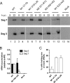
Effects of synonymous mutations on M1 and NP expression and incorporation into virions. (A) Lysates from MDCK cells infected (or mock infected) at an MOI of 1 with the indicated viruses and harvested at 24 h p.i. were analyzed by SDS-PAGE and Western blotting for NP or M1, as labeled. (B) Approximately 1 × 10⁹ virus particles from infected eggs were pelleted through a sucrose cushion, resuspended, and analyzed by SDS-PAGE and Western blotting for NP and M1. (C) The ratio of NP to M1 protein in two independently rescued egg-grown virus stocks was determined by densitometry of Western blots. Mean plus half-range values normalized with respect to those of WT virus are plotted; the bar indicates viruses with changes to conserved codons.
FIG. 5.
FIG. 5.
M2 expression in WT and mutant virus-infected cells. MDCK cells were infected with the indicated viruses (or mock infected) at an approximate MOI of 1, fixed at 8 h p.i., and stained for M2 (green) and NP (red) by immunofluorescence.
FIG. 6.
FIG. 6.
Effects of synonymous mutations on vRNA synthesis. (A) vRNA contents of total RNAs isolated at 8 or 22 h p.i. from MDCK cells infected (or mock infected) with the indicated viruses at an MOI of 0.5 were determined by reverse transcriptase primer extension with oligonucleotides specific for segments 5 and 7. (B) Segment 5 and 7 vRNA accumulation at 22 h p.i. from WT and M2 H90-V92 viruses was quantified by densitometry. Means plus SD (for five independent experiments) are plotted relative to levels in WT virus-infected samples. (C) Expression of individual vRNAs in single infected cells. Cells were infected with the indicated viruses at an empirically determined dilution that resulted in not all cells expressing NP, fixed at 12 h p.i., and stained by immunofluorescence for NP and by FISH for either vRNA segments 7 and 2 or 5 and 8 (see the legend to Fig. 9 for further details). Cells were scored for the percentage of infected cells (defined as expressing NP protein) that failed to also express one or more of the vRNA segments stained by FISH. Data are plotted as means plus SD for four experiments, two stained for segments 2 and 7 and two stained for segments 5 and 8.
FIG. 7.
FIG. 7.
EM analysis of virus budding and assembly. (A) MDCK cells were infected (or mock infected) with the indicated viruses at an MOI of 4 and fixed at 8 h p.i. before analysis by transmission EM. Bar, 500 nm. Insets show individual virions at an increased magnification. (B) Egg-grown virus was purified and examined by negative stain transmission EM. Representative virions are shown.
FIG. 8.
FIG. 8.
Effects of synonymous mutations on segment packaging. (A and C) RNAs were extracted from the allantoic fluid of infected eggs or the culture medium of infected MDCK cells, and segment 5 and 7 vRNA contents were determined by qRT-PCR. Segment concentrations were calculated with respect to plasmid standards. (A) The matching particle titers (Fig. 2) were used to calculate segment/particle ratios that were then normalized with respect to that seen for the WT virus. The mean plus half-range values for two independently rescued virus stocks are shown. (B) Egg-grown virions were pelleted from infected allantoic fluid, and approximately equal amounts of RNA (corresponding to approximately 6 × 10⁹ WT virus particles) were separated by urea-PAGE and detected by silver staining (lanes 1 to 6). Mock-infected allantoic fluid was analyzed in parallel (lane 7). (C) Plaque titer data (Fig. 2) were used to calculate the segment/PFU ratios of the indicated viruses. Values were normalized to those of the WT egg-grown virus. The means and half-ranges for two independently rescued virus stocks are shown (except for MDCK-grown WT virus, for which the mean and SD for three independent isolates are plotted).
FIG. 9.
FIG. 9.
FISH analysis of vRNA content of individual infected cells. MDCK cells were infected with the indicated viruses at an empirically determined dilution that resulted in not all cells expressing NP. Cells were fixed at 12 h p.i. and stained by immunofluorescence for NP (green) and by FISH for segments 7 (red) and 2 (magenta). Nuclei were stained with DAPI (4′,6-diamidino-2-phenylindole) (blue). Open arrowheads highlight cells expressing only NP protein, filled arrowheads show those expressing NP and a single vRNA, and arrows show cells expressing NP and both vRNAs, but with nuclear export failing to occur.

References

    1. Abramoff, M. D., P. J. Magelhaes, and S. J. Ram. 2004. Image processing with ImageJ. Biophotonics Int. 1136-42.
    1. Amorim, M. J., E. K. Read, R. M. Dalton, L. Medcalf, and P. Digard. 2007. Nuclear export of influenza A virus mRNAs requires ongoing RNA polymerase II activity. Traffic 81-11. - PubMed
    1. Bergmann, M., and T. Muster. 1996. Mutations in the nonconserved noncoding sequences of the influenza A virus segments affect viral vRNA formation. Virus Res. 4423-31. - PubMed
    1. Chao, L., T. T. Tran, and T. T. Tran. 1997. The advantage of sex in the RNA virus phi6. Genetics 147953-959. - PMC - PubMed
    1. Cheung, T. K., Y. Guan, S. S. Ng, H. Chen, C. H. Wong, J. S. Peiris, and L. L. Poon. 2005. Generation of recombinant influenza A virus without M2 ion-channel protein by introduction of a point mutation at the 5′ end of the viral intron. J. Gen. Virol. 861447-1454. - PubMed

Publication types

LinkOut - more resources