

Functional analysis of monocyte MHC class II compartments
- PMID: 18815360
- PMCID: PMC2626613
- DOI: 10.1096/fj.08-109439
Functional analysis of monocyte MHC class II compartments
Abstract
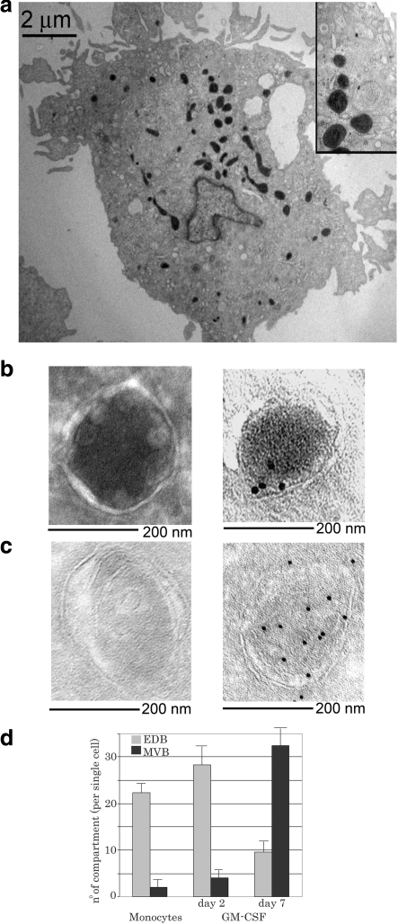
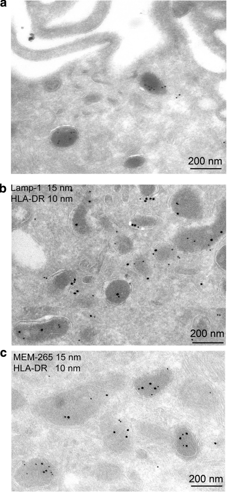
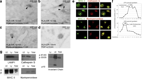
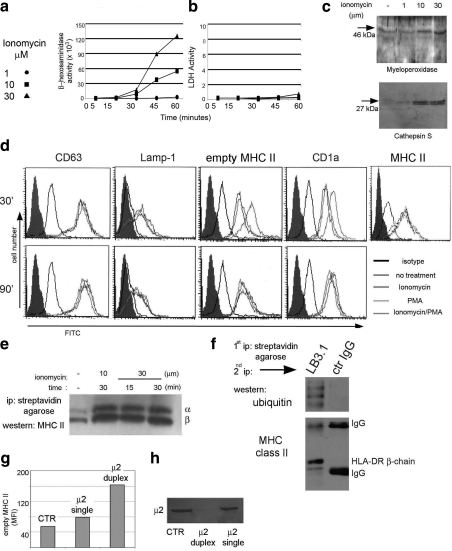
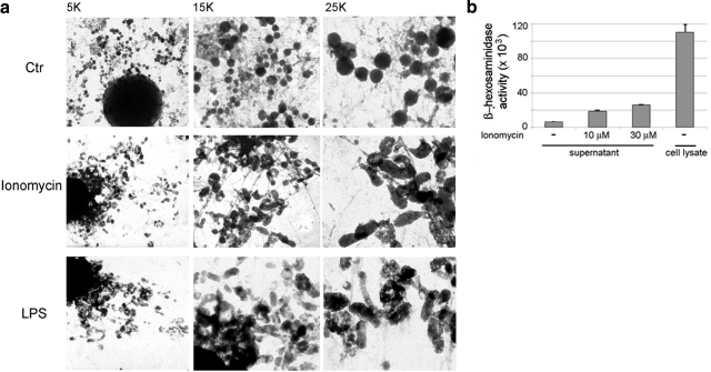
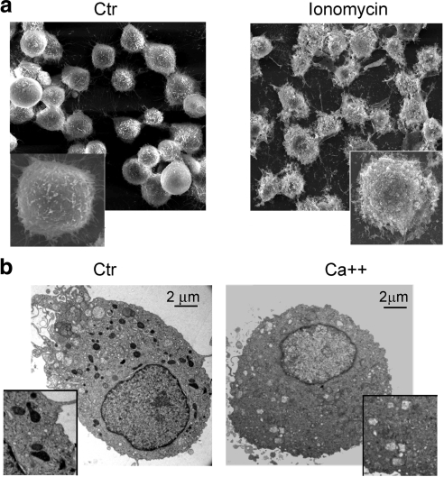
Circulating monocytes, as dendritic cell and macrophage precursors, exhibit several functions usually associated with antigen-presenting cells, such as phagocytosis and presence of endosomal/lysosomal degradative compartments particularly enriched in Lamp-1, MHC class II molecules, and other proteins related to antigen processing and MHC class II loading [MHC class II compartments (MIICs)]. Ultrastructural analysis of these organelles indicates that, differently from the multivesicular bodies present in dendritic cells, in monocytes the MIICs are characterized by a single perimetral membrane surrounding an electron-dense core. Analysis of their content reveals enrichment in myeloperoxidase, an enzyme classically associated with azurophilic granules in granulocytes and mast cell secretory lysosomes. Elevation in intracellular free calcium levels in monocytes induced secretion of beta-hexosaminidase, cathepsins, and myeloperoxidase in the extracellular milieu; surface up-regulation of MHC class II molecules; and appearance of lysosomal resident proteins. The Ca(2+)-regulated surface transport mechanism of MHC class II molecules observed in monocytes is different from the tubulovesicular organization of the multivesicular bodies previously reported in dendritic cells and macrophages. Hence, in monocytes, MHC class II-enriched organelles combine degradative functions typical of lysosomes and regulated secretion typical of secretory lysosomes. More important, Ca(2+)-mediated up-regulation of surface MHC class II molecules is accompanied by extracellular release of lysosomal resident enzymes.
Figures
Similar articles
-
MHC class II compartments in human dendritic cells undergo profound structural changes upon activation.Traffic. 2002 Dec;3(12):894-905. doi: 10.1034/j.1600-0854.2002.31205.x. Traffic. 2002. PMID: 12453152
-
3-D Structure of multilaminar lysosomes in antigen presenting cells reveals trapping of MHC II on the internal membranes.Traffic. 2004 Dec;5(12):936-45. doi: 10.1111/j.1600-0854.2004.00235.x. Traffic. 2004. PMID: 15522096
-
Identification and characterization of major histocompatibility complex class II compartments in cortical thymic epithelial cells.Anat Rec A Discov Mol Cell Evol Biol. 2003 Sep;274(1):798-806. doi: 10.1002/ar.a.10081. Anat Rec A Discov Mol Cell Evol Biol. 2003. PMID: 12923890
-
Intracellular organelles involved in antigen processing and the binding of peptides to class II MHC molecules.Semin Immunol. 1995 Dec;7(6):355-60. doi: 10.1006/smim.1995.0040. Semin Immunol. 1995. PMID: 8775461 Review.
-
MHC class II compartments: specialized organelles of the endocytic pathway in antigen presenting cells.Biol Chem. 1997 Aug;378(8):751-8. Biol Chem. 1997. PMID: 9377469 Review.
Cited by
-
Gene expression and activity of cartilage degrading glycosidases in human rheumatoid arthritis and osteoarthritis synovial fibroblasts.Arthritis Res Ther. 2009;11(3):R68. doi: 10.1186/ar2697. Epub 2009 May 14. Arthritis Res Ther. 2009. PMID: 19442276 Free PMC article.
-
Oxygen tension modulates differentiation and primary macrophage functions in the human monocytic THP-1 cell line.PLoS One. 2013;8(1):e54926. doi: 10.1371/journal.pone.0054926. Epub 2013 Jan 23. PLoS One. 2013. PMID: 23355903 Free PMC article.
-
Fingolimod Suppresses the Proinflammatory Status of Interferon-γ-Activated Cultured Rat Astrocytes.Mol Neurobiol. 2019 Sep;56(9):5971-5986. doi: 10.1007/s12035-019-1481-x. Epub 2019 Jan 30. Mol Neurobiol. 2019. PMID: 30701416
-
Low Dose Radiation Therapy Induces Long-Lasting Reduction of Pain and Immune Modulations in the Peripheral Blood - Interim Analysis of the IMMO-LDRT01 Trial.Front Immunol. 2021 Oct 12;12:740742. doi: 10.3389/fimmu.2021.740742. eCollection 2021. Front Immunol. 2021. PMID: 34712229 Free PMC article. Clinical Trial.
-
A human promyelocytic-like population is responsible for the immune suppression mediated by myeloid-derived suppressor cells.Blood. 2011 Aug 25;118(8):2254-65. doi: 10.1182/blood-2010-12-325753. Epub 2011 Jul 6. Blood. 2011. PMID: 21734236 Free PMC article.
References
-
- Geissmann F, Jung S, Littman D R. Blood monocytes consist of two principal subsets with distinct migratory properties. Immunity. 2003;19:71–82. - PubMed
-
- Randolph G J, Inaba K, Robbiani D F, Steinman R M, Muller W A. Differentiation of phagocytic monocytes into lymph node dendritic cells in vivo. Immunity. 1999;11:753–761. - PubMed
-
- Potolicchio I, Chitta S, Xu X, Fonseca D, Crisi G, Horejsi V, Strominger J L, Stern L J, Raposo G, Santambrogio L. Conformational variation of surface class II MHC proteins during myeloid dendritic cell differentiation accompanies structural changes in lysosomal MIIC. J Immunol. 2005;175:4935–4947. - PubMed
-
- Kleijmeer M J, Raposo G, Geuze H J. Characterization of MHC class II compartments by immunoelectron microscopy. Methods. 1996;10:191–207. - PubMed
-
- Stern L J, Potolicchio I, Santambrogio L. MHC class II compartment subtypes: structure and function. Curr Opin Immunol. 2006;18:64–69. - PubMed
Publication types
MeSH terms
Substances
LinkOut - more resources
Full Text Sources
Research Materials
Miscellaneous
