. 2008 Sep 30;105(39):14981-6.
doi: 10.1073/pnas.0803988105. Epub 2008 Sep 24.

Chemical targeting of the innate antiviral response by histone deacetylase inhibitors renders refractory cancers sensitive to viral oncolysis

Affiliations

Chemical targeting of the innate antiviral response by histone deacetylase inhibitors renders refractory cancers sensitive to viral oncolysis

Thi Lien-Anh Nguyên et al. Proc Natl Acad Sci U S A. .

Abstract

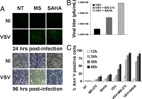
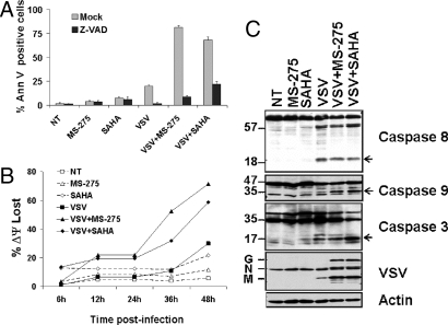
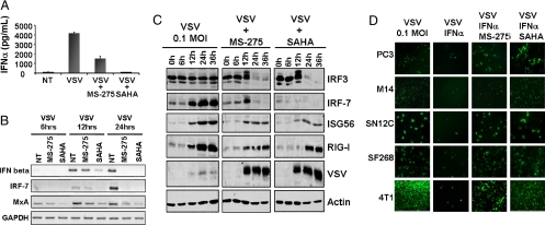
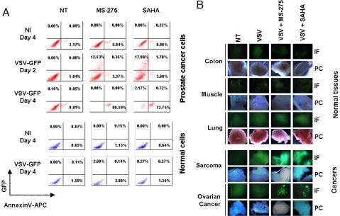
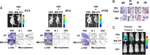
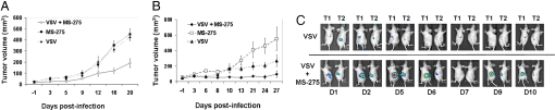
Intratumoral innate immunity can play a significant role in blocking the effective therapeutic spread of a number of oncolytic viruses (OVs). Histone deacetylase inhibitors (HDIs) are known to influence epigenetic modifications of chromatin and can blunt the cellular antiviral response. We reasoned that pretreatment of tumors with HDIs could enhance the replication and spread of OVs within malignancies. Here, we show that HDIs markedly enhance the spread of vesicular stomatitis virus (VSV) in a variety of cancer cells in vitro, in primary tumor tissue explants and in multiple animal models. This increased oncolytic activity correlated with a dampening of cellular IFN responses and augmentation of virus-induced apoptosis. These results illustrate the general utility of HDIs as chemical switches to regulate cellular innate antiviral responses and to provide controlled growth of therapeutic viruses within malignancies. HDIs could have a profoundly positive impact on the clinical implementation of OV therapeutics.


Conflict of interest statement

The authors declare no conflict of interest.

Figures

Fig. 1.
Fig. 1.
HDIs enhance VSV oncolysis in partially resistant PC3 cancer cell line. PC3 cells were either not treated (NT) or pretreated with MS-275 or SAHA for 24 h, then infected or not (NI) with VSV-Δ51-GFP. (A) Viral replication was assessed by fluorescent microscopy for GFP expression after infection with VSV at 10−4 MOI. Phase-contrast microscopy at 96 h demonstrated massive cell death in combination-treated cells. (B) Viral titers as determined by standard plaque assay after VSV infection at 0.1 MOI. (C) Induction of apoptosis was established by Annexin-V staining after VSV infection at 0.1 MOI.
Fig. 2.
Fig. 2.
VSV plus HDI combination treatment synergistically increases apoptosis in PC3 cells. PC3 cells were either not treated (NT) or pretreated with MS-275 or SAHA for 24 h then infected with VSV at 0.1 MOI. (A) Induction of apoptosis was measured by Annexin V staining in the presence or absence of the pan-caspase inhibitor Z-VADfmk. (B) Mitochondrial membrane-depolarization was assessed by JC-1 staining. (C) Caspase 8, 3, and 9 cleavage was determined 48 h after VSV infection in the presence or absence of HDIs by immunoblot of whole cell lysates. Cleaved caspase forms are indicated by open arrows.
Fig. 3.
Fig. 3.
HDIs augments VSV replication through inhibition of the IFN antiviral response. PC3 cells were either not treated (NT) or pretreated with MS-275 or SAHA for 24 h then infected with VSV at 0.1 MOI. (A) IFN-α levels in supernatants were assayed by ELISA at 24 h after infection. (B) Induction of antiviral genes IFN-beta, IRF-7 and Mxa was assessed at different times after infection by RT-PCR. NT denotes non-HDI treated cells (C) IFN-stimulated antiviral genes were analyzed by immunoblot at different times after VSV alone or VSV plus HDI treatment. (D) Different cell lines were pretreated with HDIs for 7 h and then infected with VSV-Δ51-GFP at 0.1 MOI in the presence or absence of recombinant IFNα. GFP expression was monitored 24 h after VSV inoculation.
Fig. 4.
Fig. 4.
HDI pretreatment enhances VSV oncolytic activity in primary tumor specimens. (A) Ex-vivo primary cancer or normal prostate cells were subjected to 24 h HDI pretreatment followed by VSV-Δ51GFP infection (5 MOI). VSV replication (GFP, y axis) and apoptosis induction (AnnexinV-APC staining, x axis) were determined at 2 and 4 days after infection by FACS. NT denotes non-HDI treated cells, NI denotes non-VSV-infected samples. (B) Human ex-vivo cancer or normal tissue specimens were inoculated with VSV-Δ51-GFP in the absence or presence of HDI pretreatment for 7 h. GFP expression was monitored 48 h after viral inoculation by fluorescence microscopy (IF). Phase contrast (PC) images of tissue samples are shown. NT denotes non-HDI treated, non-VSV-infected cells.
Fig. 5.
Fig. 5.
HDI treatment specifically enhances VSV-Δ51-Luc replication at the tumor site. (A) PC3, M14, and HT29 s.c. xenograft tumor models were established in nude mice and treated with a single intratumoral VSV-Δ51-Luc injection (1 × 10⁶ pfu) alone or in combination with MS-275 IP every 24 h. Viral replication at the tumor site was imaged using the IVIS system. Fluorescent orange microspheres were perfused to outline the tumor microvasculature (Microspheres). Frozen tumor sections were stained with anti-VSV antisera (α-VSV). NT = non-HDI treated, N.I. = non-VSV-infected. (B) Acetylation of histone H3 proteins was assessed in PC3 tumors by IHC at 6 h and 24 h after single IP delivery of MS-275. Skin sections were used as normal controls. (C) Transgenic mice bearing bilateral ovarian tumors were administered a single dose of VSV IP (1 × 10⁸ pfu) alone or in combination with MS-275 IP. Viral replication in live animals was assessed 48 h after viral infection by IVIS imaging.
Fig. 6.
Fig. 6.
HDI plus VSV combination treatment augments tumor-specific viral replication and significantly reduces tumor size. (A) Immunocompetent BALB/c mice bearing s.c. 4T1 tumors were treated with VSV (1 × 10⁸ pfu), alone or in combination with MS-275 IP. The efficacy of MS-275, VSV and VSV/MS-275 combination treatment in reducing tumor growth was assessed by tumor volume measurement over time. The average tumor size and standard error for each treatment group was calculated. (B) and (C) CD1 nude mice bearing SW620 tumors in hind flanks were treated with a single i.v. VSV injection (1 × 10⁷ pfu) alone or in combination with MS-275. The efficacy of MS-275, VSV and VSV/MS-275 combination treatment in reducing tumor growth was assessed by tumor volume measurement over time (B). Virus replication at the tumor site was revealed in live animals by IVIS imaging (C). Time course of treatments are schematically presented in Figs. S5 and S6.

References

    1. Bell JC. Oncolytic viruses: What's next? Curr Cancer Drug Targets. 2007;7:127–131. - PubMed
    1. Crompton AM, Kirn DH. From ONYX-015 to armed vaccinia viruses: The education and evolution of oncolytic virus development. Curr Cancer Drug Targets. 2007;7:133–139. - PubMed
    1. Kirn D, Martuza RL, Zwiebel J. Replication-selective virotherapy for cancer: Biological principles, risk management and future directions. Nat Med. 2001;7:781–787. - PubMed
    1. Parato KA, Senger D, Forsyth PA, Bell JC. Recent progress in the battle between oncolytic viruses and tumours. Nat Rev Cancer. 2005;5:965–976. - PubMed
    1. Stanford MM, McFadden G. Myxoma virus and oncolytic virotherapy: A new biologic weapon in the war against cancer. Expert Opin Biol Ther. 2007;7:1415–1425. - PubMed

Publication types

MeSH terms

LinkOut - more resources