

Lymphovascular invasion and lobular histology are associated with increased incidence of isolated tumor cells in sentinel lymph nodes from early-stage breast cancer patients
- PMID: 18815841
- PMCID: PMC4331098
- DOI: 10.1245/s10434-008-0153-2
Lymphovascular invasion and lobular histology are associated with increased incidence of isolated tumor cells in sentinel lymph nodes from early-stage breast cancer patients
Abstract
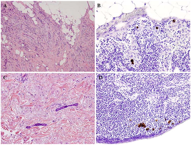
Background: Isolated tumor cells (ITC) are more likely to be identified when serial sectioning and immunohistochemical staining are used to evaluate sentinel lymph nodes (SLN). Our goal was to identify clinicopathologic features associated with ITC in patients undergoing sentinel lymph node dissection (SLND).
Methods: We reviewed clinicopathologic data for 3557 patients with no clinical evidence of lymph node metastases undergoing SLND between November 1993 and March 2007. Patients were staged according to the 6th edition of the American Joint Committee on Cancer staging system, with metastasis <or=.2 mm classified as ITC.
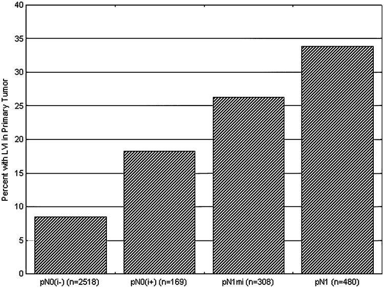
Results: A SLN was identified in 3475 patients (97.7%), including 2518 (72.4%) with negative nodes and 169 (4.9%) with ITC. A statistically significant association existed between lobular histology and the identification of ITC; 13.6% of patients with ITC had lobular histology versus 7.3% of patients with a negative SLN (P = .003). The presence of lymphovascular invasion (LVI) was also associated with ITC; 18.3% of patients with ITC had LVI in the primary tumor versus 8.5% of patients with a negative SLN (P < .001). No difference existed between patients with and without ITC with respect to T stage, grade, estrogen receptor, progesterone receptor, HER2/neu status, or biopsy method.
Conclusion: The association between ITC and LVI, a known predictor of poor outcome, suggests ITC may have clinical relevance. The relationship between lobular histology and ITC is consistent with the known pattern of lobular metastases, which frequently present as small foci requiring immunohistochemistry for detection. Longer follow-up is needed to determine whether ITC have prognostic significance.
Figures
Comment in
-
Isolated tumor cells in sentinel lymph node and clinical implications for early breast cancer.Ann Surg Oncol. 2009 Sep;16(9):2659-60; author reply 2661. doi: 10.1245/s10434-009-0455-z. Epub 2009 Jun 19. Ann Surg Oncol. 2009. PMID: 19543773 No abstract available.
-
Multidisciplinary considerations in the implementation of the findings from the American College of Surgeons Oncology Group (ACOSOG) Z0011 study: a practice-changing trial.Ann Surg Oncol. 2011 Sep;18(9):2407-12. doi: 10.1245/s10434-011-1593-7. Ann Surg Oncol. 2011. PMID: 21327455 Free PMC article. No abstract available.
References
-
- Mansel RE, Fallowfield L, Kissin M, et al. Randomized multicenter trial of sentinel node biopsy versus standard axillary treatment in operable breast cancer: the ALMANAC Trial. J Natl Cancer Inst. 2006;98:599–609. - PubMed
-
- Purushotham AD, Upponi S, Klevesath MB, et al. Morbidity after sentinel lymph node biopsy in primary breast cancer: results from a randomized controlled trial. J Clin Oncol. 2005;23:4312–21. - PubMed
-
- Veronesi U, Paganelli G, Viale G, et al. A randomized comparison of sentinel-node biopsy with routine axillary dissection in breast cancer. N Engl J Med. 2003;349:546–53. - PubMed
-
- Liberman L. Pathologic analysis of sentinel lymph nodes in breast carcinoma. Cancer. 2000;88:971–7. - PubMed
MeSH terms
Grants and funding
LinkOut - more resources
Full Text Sources
Medical
Research Materials
Miscellaneous
