

Using an amino acid fluorescence resonance energy transfer pair to probe protein unfolding: application to the villin headpiece subdomain and the LysM domain
- PMID: 18816063
- PMCID: PMC4224107
- DOI: 10.1021/bi8012406
Using an amino acid fluorescence resonance energy transfer pair to probe protein unfolding: application to the villin headpiece subdomain and the LysM domain
Abstract
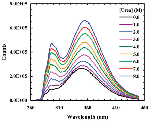
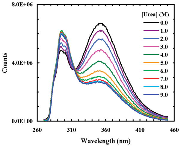
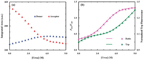
Previously, we have shown that p-cyanophenylalanine (Phe CN) and tryptophan (Trp) constitute an efficient fluorescence resonance energy transfer (FRET) pair that has several advantages over commonly used dye pairs. Here, we aim to examine the general applicability of this FRET pair in protein folding-unfolding studies by applying it to the urea-induced unfolding transitions of two small proteins, the villin headpiece subdomain (HP35) and the lysin motif (LysM) domain. Depending on whether Phe CN is exposed to solvent, we are able to extract either qualitative information about the folding pathway, as demonstrated by HP35, which has been suggested to unfold in a stepwise manner, or quantitative thermodynamic and structural information, as demonstrated by LysM, which has been shown to be an ideal two-state folder. Our results show that the unfolding transition of HP35 reported by FRET occurs at a denaturant concentration lower than that measured by circular dichroism (CD) and that the loop linking helix 2 and helix 3 remains compact in the denatured state, which are consistent with the notion that HP35 unfolds in discrete steps and that its unfolded state contains residual structures. On the other hand, our FRET results on the LysM domain allow us to develop a model for extracting structural and thermodynamic parameters about its unfolding, and we find that our results are in agreement with those obtained by other methods. Given the fact that Phe CN is a non-natural amino acid and, thus, amenable to incorporation into peptides and proteins via existing peptide synthesis and protein expression methods, we believe that the FRET method demonstrated here is widely applicable to protein conformational studies, especially to the study of relatively small proteins.
Figures


References
-
- Sapsford KE, Berti L, Igor L, Medintz IL. Materials for fluorescence resonance energy transfer analysis: Beyond traditional donor-acceptor combinations. Angew Chem, Int Ed. 2006;45:4562–4588. - PubMed
-
- Royer CA. Probing protein folding and conformational transitions with fluorescence. Chem Rev. 2006;106:1769–1784. - PubMed
-
- Tucker MJ, Oyola R, Gai F. Conformational distribution of a 14 residue peptide in solution: A fluorescence resonance energy transfer study. J Phys Chem B. 2005;109:4788–4795. - PubMed
Publication types
MeSH terms
Substances
Grants and funding
LinkOut - more resources
Full Text Sources
Other Literature Sources
