Review
. 2009 Feb;11(2):369-99.
doi: 10.1089/ars.2008.2130.

Modulation of brain hemichannels and gap junction channels by pro-inflammatory agents and their possible role in neurodegeneration

Affiliations
Review

Modulation of brain hemichannels and gap junction channels by pro-inflammatory agents and their possible role in neurodegeneration

Juan A Orellana et al. Antioxid Redox Signal. 2009 Feb.

Abstract

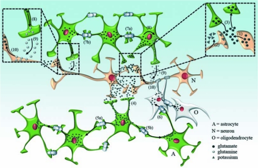
In normal brain, neurons, astrocytes, and oligodendrocytes, the most abundant and active cells express pannexins and connexins, protein subunits of two families forming membrane channels. Most available evidence indicates that in mammals endogenously expressed pannexins form only hemichannels and connexins form both gap junction channels and hemichannels. Whereas gap junction channels connect the cytoplasm of contacting cells and coordinate electric and metabolic activity, hemichannels communicate the intra- and extracellular compartments and serve as a diffusional pathway for ions and small molecules. A subthreshold stimulation by acute pathological threatening conditions (e.g., global ischemia subthreshold for cell death) enhances neuronal Cx36 and glial Cx43 hemichannel activity, favoring ATP release and generation of preconditioning. If the stimulus is sufficiently deleterious, microglia become overactivated and release bioactive molecules that increase the activity of hemichannels and reduce gap junctional communication in astroglial networks, depriving neurons of astrocytic protective functions, and further reducing neuronal viability. Continuous glial activation triggered by low levels of anomalous proteins expressed in several neurodegenerative diseases induce glial hemichannel and gap junction channel disorders similar to those of acute inflammatory responses triggered by ischemia or infectious diseases. These changes are likely to occur in diverse cell types of the CNS and contribute to neurodegeneration during inflammatory process.


Figures

FIG. 1.
FIG. 1.
Scheme showing the membrane topology of pannexin, connexin, hemichannels, and gap junction channels. Top and bottom right correspond to pannexin and connexin in a plasma membrane, respectively. Both protein types have four transmembrane domains (M1–4) with amino (—NH2) and carboxy (—COOH) termini on the cytoplasmic side, two extracellular loops (E1 and E2), and one cytoplasmic loop (CL). Top and bottom center show hemichannels formed of six pannexin or connexin subunits each. The middle center shows an aggregate of connexin gap junction channels, a section through a gap junction “plaque”, at a close contact between cells 1 and 2, as shown in the left. Each gap junction channel is formed by two hemichannels docked together (and rotated 30° with respect to one another). Each cell contributes one of hemichannels.
FIG. 2.
FIG. 2.
Cellular distribution of pannexin and connexin hemichannels and connexin gap junctions in brain cells. This figure includes only those cases in which the available information has been obtained in vivo and/or in vitro with more than one experimental approach. Homocellular (A–G) and heterocellular (H and I) connexin gap junction channels and connexin and pannexin hemichannels (A, B, and D) are indicated within the encircled regions. Heterotypic gap junction channels (i.e., with different connexins in the two hemichannels) are indicated in B and I. Neuron (N), astrocyte (A), oligodendrocyte (O), microglia (M), endothelial cell (EC), ependimocyte (E), and meningeal cell (MC) are denoted in panels A–I.
FIG. 3.
FIG. 3.
Role of astroglial gap junction communication in K+ spatial buffering and the tripartite chemical synapse. Glutamate released from presynaptic neurons (1) binds to ionotropic glutamate receptors, triggering a postsynaptic potential in the postsynaptic neuron and promoting the K+ release (2) during repolarization that is more prominent if action potentials are elicited (Box, enlarged in the right inset). Astrocytes surrounding the synapses take up glutamate (3) through EAAT1 and EAAT2 transporters. During high rates of neuronal activity, K+ accumulates in the extracellular space, and then is taken up by astrocytes (4) through at least inwardly rectifying potassium channels and Na+/K+-pumps. K+ that accumulates inside astrocytes diffuses to neighboring astrocytes (5a to 5b, follow arrows in astrocytes at the bottom) and oligodendrocytes (6) via gap junction channels, a process termed “spatial buffering”. Spatial buffering is contributed to by depolarization in regions of K+ accumulation; the increased positivity causes a current to flow out through membrane that is less depolarized. This outward flow is carried by K+, which is also the major charge carrier in the cytoplasm. Similarly, glutamate taken up by astrocytes diffuses (7a to 7b, follow arrows in astrocytes at the top) to neighboring astrocytes through gap junction channels. The glutamate is metabolized to glutamine (8) by glutamine synthetase and released to the extracellular milieu from which it is taken up by neurons (9) (Box, enlarged in the left inset). In neurons, glutamine is transformed to the neurotransmitter, glutamate (or GABA) (10). (For interpretation of the references to color in this figure legend, the reader is referred to the web version of this article at www.liebertonline.com/ars).
FIG. 4.
FIG. 4.
Role of connexins in neuron/glial metabolic coupling. (1) Under normal conditions, endothelial cells of the blood brain barrier (BBB) take up blood-borne glucose and lactate through GLUT-1 and monocarboxylate transporters, respectively. Both lactate (2) or glucose (3) may diffuse through gap junction channels between adjacent endothelial cells. Both metabolites are eventually taken up by astrocytic endfeet (4) or released to the interstitial space (5). Glucose (6) and lactate (7) can diffuse through astrocytes and their gap junctions with neighboring astrocytes to reach relatively distant areas. Glucose can be metabolized to lactate by astrocytes (8), and both can be released into the extracellular space and taken up by neurons (9). Astrocytes may transfer lactate and/or glucose to oligodendrocytes through heterocellular gap junction channels between them (10). (For interpretation of the references to color in this figure legend, the reader is referred to the web version of this article at www.liebertonline.com/ars).
FIG. 5.
FIG. 5.
Connexin based channels in brain cells during bacterial meningitis. During bacterial infection blood levels of cytokine (e.g., TNF-α and IL-β) are elevated. Both, TNF-α and IL-1β enhance hemichannel activity (1) of brain endothelial cells. Furthermore, these cytokines induce BBB discontinuity favoring bacterial extravasation (2). Once in the interstitium, bacteria and their extracellular wall components such as LPS and PGN are recognized by microglia (3) which are activated and release cytokines that further activate them (reciprocal arrows). ATP released via hemichannels from microglia (4) promotes microglial migration from less affected regions. Activated microglia can also release glutamate through hemichannels and oxygen- and nitrogen-derived free radicals that are neurotoxic (5). The enhanced hemichannel activity of activated astrocytes induces neuronal damage through the release of neurotoxic and/or inflammatory compounds such as glutamate and PGE2 (6). These compounds may also increase the activity of neuronal pannexin hemichannels (7), causing electrochemical imbalance and Ca2+ overload in neurons. In contrast to increased opening of hemichannels, astroglial gap junction communication is reduced (8) (depicted in the figure as reduction in channel number, but could also be the consequence of a reduction in permeability or open probability), impairing glutamate and K+ spatial buffering, which enhances neuronal susceptibility to insults. Bacterial meningitis can also induce demyelination (9), possibly via microglial cytokine release. Severe inflammation induces recruitment of leucocytes (10) to the infected loci. Gap junction communication between leucocytes and endothelial cells (11) may contribute to stronger heterocellular adhesion and allow transfer of signals that regulate leucocyte diapedesis across the endothelium (12). Activated microglia may perform antigen cross-presentation interaction with infiltrating leucocytes in which gap junctions between them may play an important role (13). Direct microglial interaction with LPS or PGN induces gap junction communication between microglia which can coordinate microglial function (14). (For interpretation of the references to color in this figure legend, the reader is referred to the web version of this article at www.liebertonline.com/ars).
FIG. 6.
FIG. 6.
Microglial conditioned medium enhances dye uptake in RBE4 cells, a cell line derived from brain endothelial cells. In RBE4 cells, ethidium (Et) (5 μM) uptake was measured every 30 s as fluorescence emission of Et bound to DNA (518 nm, intensity in arbitrary units, AU). (A) Fluorescence micrograph of Et uptake taken after 10 min under control conditions shows little uptake. (B) After 24 h of incubation with medium conditioned by microglia treated for 6 h with LPS, uptake is increased after the same exposure to Et (10 min). (C) Time lapse measurements of Et uptake in RBE4 cells under control conditions (white circles) and after 24 h of exposure to the conditioned medium of LPS treated microglia (black circles). Under both conditions, Et uptake was blocked by La3+ (200 μM) applied after ~11 min. (D) Averaged data normalized to control (dashed line) of Et uptake rate of RBE4 cells treated for 3, 12, or 24 h with medium conditioned by microglia treated with LPS. Bar = 10 μm.
FIG. 7.
FIG. 7.
Conditioned medium from microglia treated with LPS induces Cx43 hemichannel activity in cortical astrocytes. (A) Voltage ramps from −80 to 0 mV, 3 s in duration, were applied during whole cell recording. The ramp was initiated by a transition from 0 to −80 mV. (B) Under control conditions, no hemichannel openings were observed, although there was some low level activity at the more negative potentials. (C) In astrocytes treated for 24 h with medium conditioned by microglia treated with LPS, an obvious increase in channel openings was observed. The boxed region in C is enlarged in the inset and shows ~220 pS transitions between closed and open states; this conductance is characteristic of Cx43 hemichannels.
FIG. 8.
FIG. 8.
Three mechanisms of death amplification. (A) Initially, a brain injury produced by ischemia, infection, or necrosis affecting astrocytes (green), neurons (orange), or resting microglia (blue), could start a wave of death propagated (yellow arrows) and amplified through diffusible toxins and molecules (e.g., Ca2+, NO, superoxide ion, peroxinitrite, glutamate, and NAD+) present in high concentration in injured cells (depicted in the figure as dark-colored cells). These molecules could be transferred through connexin gap junctions and connexin and pannexin hemichannels from injured cells (less and more affected cells in gray and black, respectively) to healthier cells. (B) Later, a second wave of death (yellow arrows) may be mediated by microglial cells overactivated by ATP and cytokines released by injured cells. (C) Still later inflammation-induced edema that reduces tissue perfusion could worsen the inflammatory response, recruiting leucocytes and increasing the extent of the lesion. (For interpretation of the references to color in this figure legend, the reader is referred to the web version of this article at www.liebertonline.com/ars).

References

    1. A novel gene containing a trinucleotide repeat that is expanded and unstable on Huntington's disease chromosomes. The Huntington's Disease Collaborative Research Group. Cell. 1993;72:971–983. - PubMed
    1. Aguado F. Ballabriga J. Pozas E. Ferrer I. TrkA immunoreactivity in reactive astrocytes in human neurodegenerative diseases and colchicine-treated rats. Acta Neuropathol. 1998;96:495–501. - PubMed
    1. Ahmad AS. Zhuang H. Echeverria V. Dore S. Stimulation of prostaglandin EP2 receptors prevents NMDA-induced excitotoxicity. J Neurotrauma. 2006;23:1895–1903. - PubMed
    1. Ahmad S. Evans WH. Post-translational integration and oligomerization of connexin 26 in plasma membranes and evidence of formation of membrane pores: implications for the assembly of gap junctions. Biochem J. 2002;365:693–699. - PMC - PubMed
    1. Aizenman E. Lipton SA. Loring RH. Selective modulation of NMDA responses by reduction and oxidation. Neuron. 1989;2:1257–1263. - PubMed

Publication types

LinkOut - more resources