

Four-dimensional transcatheter intraarterial perfusion (TRIP)-MRI for monitoring liver tumor embolization in VX2 rabbits
- PMID: 18816818
- PMCID: PMC2917606
- DOI: 10.1002/mrm.21678
Four-dimensional transcatheter intraarterial perfusion (TRIP)-MRI for monitoring liver tumor embolization in VX2 rabbits
Abstract
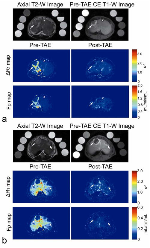
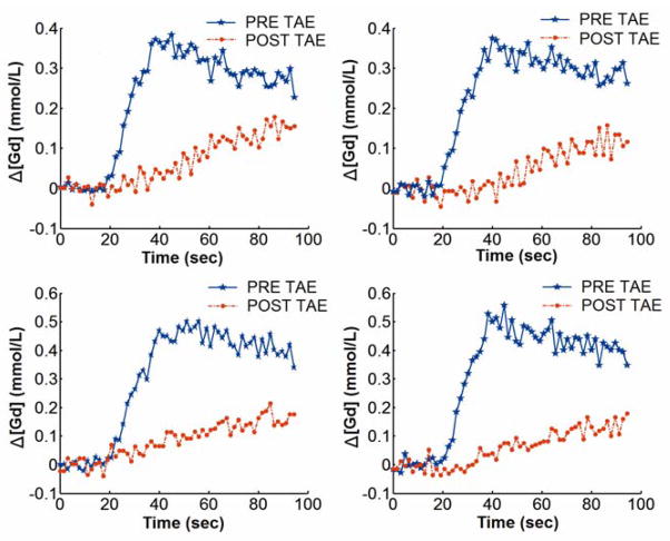
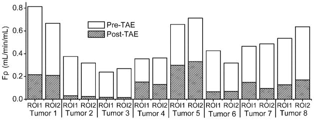
Transcatheter intraarterial perfusion (TRIP)-MRI is an intraprocedural technique to iteratively monitor liver tumor perfusion changes during transcatheter arterial embolization (TAE) and chemoembolization (TACE). However, previous TRIP-MRI approaches using two-dimensional (2D) T(1)-weighted saturation-recovery gradient-recalled echo (GRE) sequences provided only limited spatial coverage and limited capacity for accurate perfusion quantification. In this preclinical study, a quantitative 4D TRIP-MRI technique (serial iterative 3D volumetric perfusion imaging) with rigorous radiofrequency (RF) B(1) field calibration and dynamic tissue longitudinal relaxation rate R(1) measurement is presented for monitoring intraprocedural liver tumor perfusion during TAE. 4D TRIP-MRI and TAE were performed in five rabbits with eight VX2 liver tumors (N = 8). After B(1) calibrated baseline and dynamic R(1) quantification, subsequent tissue contrast agent concentration time curves were derived. A single-input flow-limited pharmacokinetic model and peak gradient method were applied for perfusion analysis. The perfusion Frho reduced significantly from pre-TAE 0.477 (95% confidence interval [CI]: 0.384-0.570) to post-TAE 0.131 (95% CI: 0.080-0.183 ml/min/ml, P < 0.001).
(c) 2008 Wiley-Liss, Inc.
Figures
References
-
- Llovet JM, Real MI, Montana X, Planas R, Coll S, Aponte J, Ayuso C, Sala M, Muchart J, Sola R, Rodes J, Bruix J. Arterial embolisation or chemoembolisation versus symptomatic treatment in patients with unresectable hepatocellular carcinoma: a randomised controlled trial. Lancet. 2002;359(9319):1734–1739. - PubMed
-
- Lewandowski RJ, Wang D, Gehl J, Atassi B, Ryu RK, Sato K, Nemcek AA, Jr, Miller FH, Mulcahy MF, Kulik L, Larson AC, Salem R, Omary RA. A comparison of chemoembolization endpoints using angiographic versus transcatheter intraarterial perfusion/MR imaging monitoring. J Vasc Interv Radiol. 2007;18(10):1249–1257. - PubMed
-
- Wang D, Bangash AK, Rhee TK, Woloschak GE, Paunesku T, Salem R, Omary RA, Larson AC. Liver tumors: monitoring embolization in rabbits with VX2 tumors--transcatheter intraarterial first-pass perfusion MR imaging. Radiology. 2007;245(1):130–139. - PubMed
-
- Virmani S, Wang D, Harris KR, Ryu RK, Sato KT, Lewandowski RJ, Nemcek AA, Jr, Szolc-Kowalska B, Woloschak G, Salem R, Larson AC, Omary RA. Comparison of transcatheter intraarterial perfusion MR imaging and fluorescent microsphere perfusion measurements during transcatheter arterial embolization of rabbit liver tumors. J Vasc Interv Radiol. 2007;18(10):1280–1286. - PubMed
-
- Larson AC, Wang D, Atassi B, Sato KT, Ryu RK, Lewandowski RJ, Nemcek AA, Jr, Mulcahy MF, Kulik LM, Miller FH, Salem R, Omary RA. Transcatheter intraarterial perfusion: MR monitoring of chemoembolization for hepatocellular carcinoma--feasibility of initial clinical translation. Radiology. 2008;246(3):964–971. - PubMed
MeSH terms
Grants and funding
LinkOut - more resources
Full Text Sources
Other Literature Sources
Medical
Miscellaneous
