. 2008 Oct;237(10):2820-9.
doi: 10.1002/dvdy.21703.

Dynamic GATA6 expression in primitive endoderm formation and maturation in early mouse embryogenesis

Affiliations

Dynamic GATA6 expression in primitive endoderm formation and maturation in early mouse embryogenesis

Kathy Q Cai et al. Dev Dyn. 2008 Oct.

Abstract

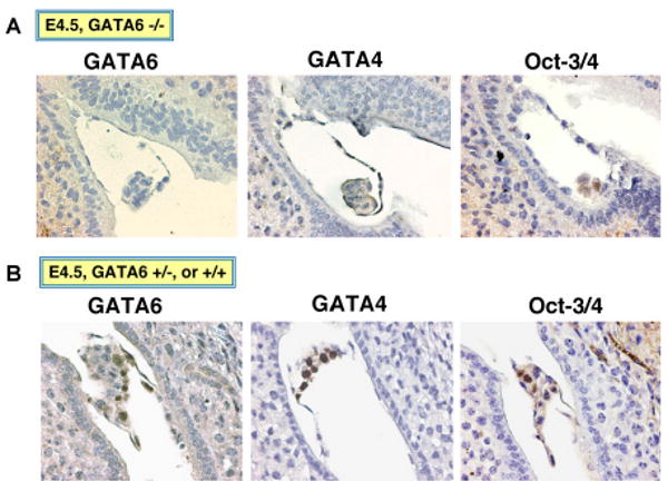
The derivation of the primitive endoderm layer from the pluripotent cells of the inner cell mass is one of the earliest differentiation and morphogenic events in embryonic development. GATA4 and GATA6 are the key transcription factors in the formation of extraembryonic endoderms, but their specific contribution to the derivation of each endoderm lineage needs clarification. We further analyzed the dynamic expression and mutant phenotypes of GATA6 in early mouse embryos. GATA6 and GATA4 are both expressed in primitive endoderm cells initially. At embryonic day (E) 5.0, parietal endoderm cells continue to express both GATA4 and GATA6; however, visceral endoderm cells express GATA4 but exhibit a reduced expression of GATA6. By and after E5.5, visceral endoderm cells no longer express GATA6. We also found that GATA6 null embryos did not form a morphologically recognizable primitive endoderm layer, and subsequently failed to form visceral and parietal endoderms. Thus, the current study establishes that GATA6 is essential for the formation of primitive endoderm, at a much earlier stage then previously recognized, and expression of GATA6 discriminates parietal endoderm from visceral endoderm lineages.


Figures

Fig. 1
Fig. 1
Expression of GATA4 and GATA6 in immediately implanted blastocysts and early embryos. AC: Fixed and paraffin-embedded uterine horns containing embryonic day (E) 4.5 (A), E4.75 (B), and E5.0 (C) embryos from timed matings of wild-type mice were sectioned through and adjacent slides were immunostained for GATA4 and GATA6. Representative stainings are shown at ×400.
Fig. 2
Fig. 2
Exclusive expression of GATA6 in parietal but not visceral endoderm in mouse embryos. Fixed and paraffin-embedded uterine horns containing embryonic day (E) 5.5, E6.0, and E7.0 embryos from timed matings of wild-type mice were sectioned and adjacent slides were used for immunostaining. Representative images are shown. A: An E5.5 embryo stained for GATA6, GATA4, and laminin, showing at ×200. B: An E6.0 embryo stained for GATA6 and GATA4, showing at ×200. C: An E7.0 embryo stained for GATA6, GATA4, Dab2, and laminin, showing at ×100. Arrow indicates visceral endoderm and arrowhead indicates parietal endoderm cells.
Fig. 3
Fig. 3
Derivation of GATA6- and/or GATA4-positive cells following the differentiation of embryonic stem Cells in monolayer cultures. Wild-type embryonic stem (ES) cells were cultured in the presence of 1 μM retinoic acid for 4 days on slide chambers. The cells were processed for immunofluorescence staining with both anti-GATA4 and anti-GATA6 antibodies. DAPI (′,6-diamidine-2-phenylidole-dihydrochloride) was used to stain the nucleus. Two representative fields are shown. Arrows indicate examples of cells that are positive for GATA4 but negative for GATA6.
Fig. 4
Fig. 4
Derivation of GATA6- and/or GATA4-positive Cells following the differentiation of embryonic stem (ES) cells in embryoid bodies. Embryoid bodies were formed following aggregation of wild-type ES cells in suspension culture for 7 days. The embryoid bodies were harvested and subjected to immunostaining. A: A representative section showing double staining for GATA4 and GATA6 immunofluorescence. DAPI was used to stain nucleus. A fraction of these endoderm cells show GATA4 staining but little or no GATA6 expression (arrows). B: Adjacent sections of embryoid bodies were stained for GATA6 and PAS, which is used to stain glycoproteins of the basement membranes. An arrow indicates presumptive visceral endoderm cells with abundant apical vacuoles, and the arrowhead indicates parietal endoderm cells adhered to PAS-stained basement membrane.
Fig. 5
Fig. 5
Endoderm differentiation of GATA6-dificient embryonic stem (ES) cells. Wild-type and GATA6 (−/−) ES cells transfected with GATA4 expression plasmids (+ GATA4) or control vectors, were cultured in monolayer or suspension (Sph) as spheroids/embryoid bodies, and/or were differentiated by addition of retinoic acid (+ RA) for 7 days. A,B: The cells were harvested and subjected to Western blot analysis using an Odysse Infrared Imaging system from LI-COR (A), or immunofluorescence staining (B). In (B), immunofluorescence staining of GATA4 indicates the transfected cells and Dab2 staining is a marker of endoderm differentiation.
Fig. 6
Fig. 6
Failure of extraembryonic endoderm formation in GATA6-deficient embryos. embryonic day (E) 5.5 and E6.5 embryos from timed matings of GATA6 (+/−) mice were analyzed by histology and immunostaining. Adjacent sections were stained for GATA4, GATA6, Oct-3/4, and Dab2. Embryos that are GATA6 negative were identified as putative GATA6 (−/−).A: An E5.5 embryo stained for GATA6, GATA4, and Oct-3/4 was identified as GATA6 (−/−).B: A presumably GATA6 (−/−) E6.5 embryo is shown for hematoxylin–eosin, GATA6, GATA4, and Dab2 staining.
Fig. 7
Fig. 7
Absence of primitive endoderm in embryonic day (E) 4.5 GATA6-deficient embryos. E4.5 embryos from timed-matings of GATA6 (+/−) mice were analyzed by histology and immunostaining. Adjacent sections were stained for GATA4, GATA6, and Oct-3/4. A: An example of a presumptive GATA6 (−/−) E4.5 embryo is shown for GATA6, GATA4, and Oct-3/4 staining. The absence of GATA6 staining, GATA4 staining, and the abnormal morphology were used to denote the potential GATA6 null genotype of the embryo. B: In the same uteri, additional embryos show GATA4- and GATA6-positive staining that was determined to be GATA6 (+/+) or (+/−).
Fig. 8
Fig. 8
Detection of variable GATA6 expression in visceral endoderm cells of embryonic day (E) 5.0 embryos. E5.0 embryos from timed matings of wild-type mice were analyzed by histology and immunostaining. Sequential sections stained for GATA4, GATA6, laminin, and Dab2 were shown. GATA6 staining varies in individual visceral endoderm cells. An arrow shows a visceral endoderm cell stained positive for GATA6, and arrowhead indicates a visceral endoderm cell with weak GATA6 staining.
Fig. 9
Fig. 9
A model for lineage derivation in postimplanted blastocysts. Schematic illustration shows a model for the lineage derivation in postimplantation blastocysts. It is hypothesized that induction of GATA4 and GATA6 expression leads to the origination of primitive endoderm in the inner cell mass (ICM), and subsequently the cells are sorted to the surface to form the primitive endoderm structure. Subsequently, a fraction of the primitive endoderm cells migrate to cover the blastocoel and differentiate into parietal endoderm, which remain positive for both GATA4 and GATA6. The remaining primitive endoderm cells covering the ICM gradually lose GATA6 expression and convert to visceral endoderm cells that are positive for GATA4 but negative for GATA6. GATA6 is essential for differentiation of the primitive endoderm lineage. Suppression of GATA6 occurs in the maturation of primitive endoderm into visceral endoderm cells.

References

    1. Arceci RJ, King AA, Simon MC, Orkin SH, Wilson DB. Mouse GATA-4: a retinoic acid-inducible GATA-binding transcription factor expressed in endodermally derived tissues and heart. Mol Cell Biol. 1993;13:2235–2246. - PMC - PubMed
    1. Beddington RS, Robertson EJ. Axis development and early asymmetry in mammals. Cell. 1999;96:195–209. - PubMed
    1. Bielinska M, Wilson DB. Induction of yolk sac endoderm in GATA-4-deficient embryoid bodies by retinoic acid. Mech Dev. 1997;65:43–54. - PubMed
    1. Capo-Chichi CD, Rula ME, Smedberg JL, Vanderveer L, Parmacek MS, Morrisey EE, Godwin AK, Xu XX. Perception of differentiation cues by GATA factors in primitive endoderm lineage determination of mouse embryonic stem cells. Dev Biol. 2005;286:574–586. - PubMed
    1. Chazaud C, Yamanaka Y, Pawson T, Rossant J. Early lineage segregation between epiblast and primitive endoderm in mouse blastocysts through the Grb2-MAPK pathway. Dev Cell. 2006;10:615–624. - PubMed

Publication types

Substances

LinkOut - more resources