Review
. 2009:27:29-60.
doi: 10.1146/annurev.immunol.24.021605.090616.

Structural biology of shared cytokine receptors

Affiliations
Review

Structural biology of shared cytokine receptors

Xinquan Wang et al. Annu Rev Immunol. 2009.

Abstract

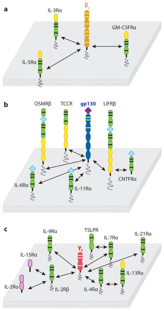
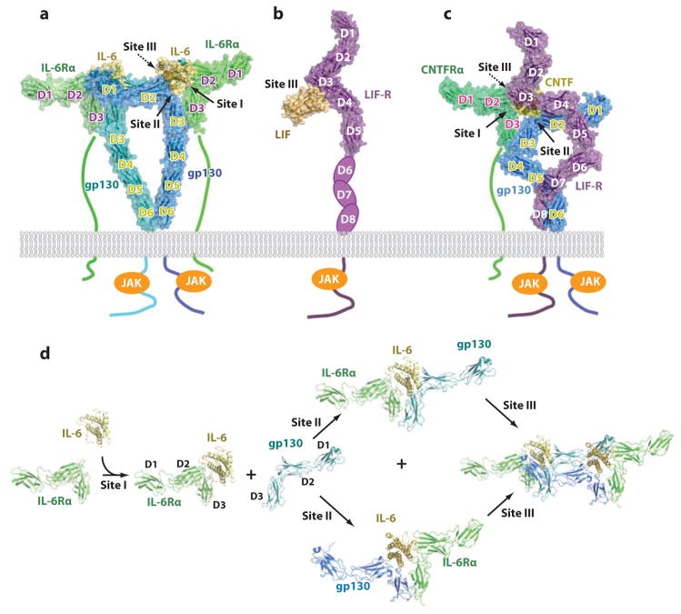
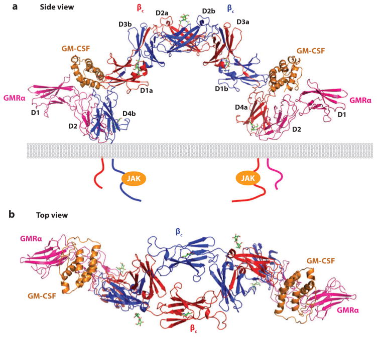
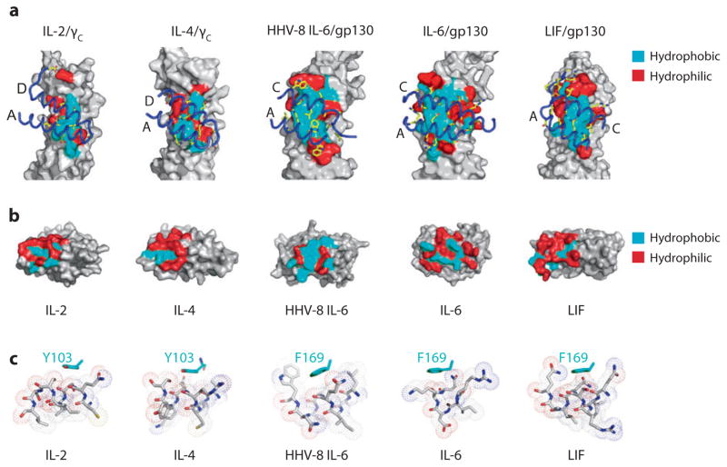
Recent structural information for complexes of cytokine receptor ectodomains bound to their ligands has significantly expanded our understanding of the macromolecular topology and ligand recognition mechanisms used by our three principal shared cytokine signaling receptors-gp130, gamma(c), and beta(c). The gp130 family receptors intricately coordinate three structurally unique cytokine-binding sites on their four-helix bundle cytokine ligands to assemble multimeric signaling complexes. These organizing principles serve as topological blueprints for the entire gp130 family of cytokines. Novel structures of gamma(c) and beta(c) complexes show us new twists, such as the use of a nonstandard sushi-type alpha receptors for IL-2 and IL-15 in assembling quaternary gamma(c) signaling complexes and an antiparallel interlocked dimer in the GM-CSF signaling complex with beta(c). Unlike gp130, which appears to recognize vastly different cytokine surfaces in chemically unique fashions for each ligand, the gamma(c)-dependent cytokines appear to seek out some semblance of a knobs-in-holes shape recognition code in order to engage gamma(c) in related fashions. We discuss the structural similarities and differences between these three shared cytokine receptors, as well as the implications for transmembrane signaling.


Figures

Figure 1
Figure 1
Diversity of shared cytokine-receptor interactions. Shared cytokine receptors γc (a), gp130 (b), and βc (c) are represented schematically on a cell membrane. The respective interacting cytokines with known three-dimensional structures are shown with cylinder representations of the four-helix bundles. (Abbreviations: LIF, leukemia inhibitory factor; OSM, oncostatin-M; CNTF, cillary neurotrophic factor; CLC, cardiotrophin-like cytokine; CHR, cytokine-binding homology region; HHV-8, human herpes virus; GM-CSF, granulocyte-macrophage colony-stimulating factor.)
Figure 2
Figure 2
Diversity of shared receptor-receptor interactions. Shared cytokine receptors βc (a), gp130 (b), and γc (c) and their various receptor partners are depicted. These complexes are formed by the combination of ligand-specific α and/or β receptors with shared cytokine receptors. (Abbreviations: TCCR, T cell cytokine receptor; TSLPR, thymic stromal-derived lymphopoietin receptor.)
Figure 3
Figure 3
Structures of human IL-6/IL-6Rα/gp130 hexameric complex (a), mouse LIFR in complex with human LIF (b), and human CNTF/CNTFRα/LIFR/gp130 (c) assembled from known crystallographic, biochemical, and electron microscopic data. In (a) the model shown was derived from the crystal structure of the IL-6 hexamer headpiece (12) together with the single-particle reconstruction of the entire extracellular complex (72). IL-6 signaling is mediated through homodimerization of gp130 in a symmetric hexameric arrangement with a nonsignaling IL-6Rα receptor. In (b) the model shown derives from the 4 Å crystal structure of the LIF/LIFR complex (14) missing the membrane-proximal domains that are depicted as cartoons. In (c), the quaternary LIFR/gp130/CNTF/CNTFRα complex is derived from a combination of the crystal structures of LIF/gp130 (13), CNTF (163), and a single-particle reconstruction of the entire quaternary complex (76). CNTF signals through the asymmetric heterodimerization of gp130 and LIFR and the nonsignaling CNTFα receptor. In panel (d), the assembly pathway for IL-6 signaling is depicted as elucidated from References , . IL-6 first engages IL-6Rα through a site I interaction to form a composite interface (site II) that recruits gp130. This trimeric structure can then engage a second trimer through two site III interfaces to form a productive signaling complex.
Figure 4
Figure 4
The 2:2:2 GM-CSF/GMRα/βc complex viewed from the side (a) and from the top (b) (18). GMRα engages the cytokine GM-CSF via a canonical site I interaction, whereas the βc receptor engages site II on GM-CSF by using a composite cytokine-binding homology region (CHR) interface generated by domain 1 (D1) of one βc subunit and domain 4 (D4) of the second βc.
Figure 5
Figure 5
Extracellular complex structures of IL-2/IL-2Rα/IL-2Rβ/γc (a) (15), IL-4/IL-4R α/γc (b), IL-4/IL-4Rα/IL-13Rα1 (c), and IL-13/IL-4Rα/IL-13Rα1 (d) (17) extracellular signaling complexes depicted on a cell membrane.
Figure 6
Figure 6
IL-2/IL-2Rα and IL-15/IL-15Rα binary complexes. (a) IL-2Rα is composed of two sushi modules (D1 and D2) that swap opposing β-strands, forming a noncanonical sushi fold topology (53). (b) IL-2Rα and IL-15Rα contact the dorsal surface of IL-2 and IL-15, respectively, with respect to the membrane (130, 131). (c) IL-2 quaternary complex and modeled IL-15 quaternary complexes. IL-2Rα presents IL-2 in cis, whereas IL-15Rα presents IL-15 in cis or trans.
Figure 7
Figure 7
The sequential assembly pathways of the IL-2/IL-2Rα/IL-2Rβ/γc quaternary (a), IL-4/IL-4Rα/γc (b), IL-4/IL-4Rα/IL-13Rα1 (c), and IL-13/IL-4Rα/IL-13Rα1 (d) ternary complexes. See Table 1 for the interaction affinity and thermodynamic parameters of each binding site.
Figure 8
Figure 8
Specific versus degenerate binding sites I and IIa in the IL-2/IL-2Rα/IL-2Rβ/γc and IL-4/IL-4Rα/γc complexes. (a) Left panel shows the binding site I between IL-2 helices A and C and IL-2Rβ loops in the elbow region. The corresponding binding site I in the IL-4 ternary complex is shown in right panel. (b) Binding site IIa in the IL-2 quaternary complex (γc/IL-2) and in the IL-4 ternary complex (γc/IL-4) are shown in left and right panels, respectively.
Figure 9
Figure 9
Cross-reactivity of the cytokine-binding homology region (CHR) of γc and gp130. (a) Contact surface of γc and gp130 when bound to IL-2, IL-4, HHV-8 IL-6, IL-6, and LIF. Hydrophobic residues are colored as blue surface area and hydrophilic residues as red surface area. The helices and the contacting residues for each of the cytokines are shown docked onto the surface of the receptor. (b) Surface representations showing the contact surface of each cytokine. Note that IL-2 and IL-4 have a similar distribution of hydrophobic and hydrophilic residues in the contact area. For gp130 family cytokines, HHV-8 IL-6 has primarily hydrophobic contact surface area. The contact area on IL-6 is more polar, and LIF has the most significant hydrophilic contact surface. (c) The packing environment of the common binding epitope residues Tyr-103 (γc) and Phe-169 (gp130) against the cytokines.

References

    1. Garcia KC, editor. Advances in Protein Chemistry. Vol. 68. London/San Diego: Academic; 2004. p. 506.
    1. Dechant G, Barde YA. The neurotrophin receptor p75(NTR): novel functions and implications for diseases of the nervous system. Nat Neurosci. 2002;5:1131–36. - PubMed
    1. Airaksinen MS, Saarma M. The GDNF family: signalling, biological functions and therapeutic value. Nat Rev Neurosci. 2002;3:383–94. - PubMed
    1. Rudolph MG, Stanfield RL, Wilson IA. How TCRs bind MHCs, peptides, and coreceptors. Annu Rev Immunol. 2006;24:419–66. - PubMed
    1. Strong RK. Asymmetric ligand recognition by the activating natural killer cell receptor NKG2D, a symmetric homodimer. Mol Immunol. 2002;38:1029–37. - PubMed

Publication types

MeSH terms

LinkOut - more resources