. 2008 Oct 14;18(19):1476-82.
doi: 10.1016/j.cub.2008.08.013. Epub 2008 Sep 25.

3' UTRs are the primary regulators of gene expression in the C. elegans germline

Affiliations

3' UTRs are the primary regulators of gene expression in the C. elegans germline

Christopher Merritt et al. Curr Biol. .

Abstract

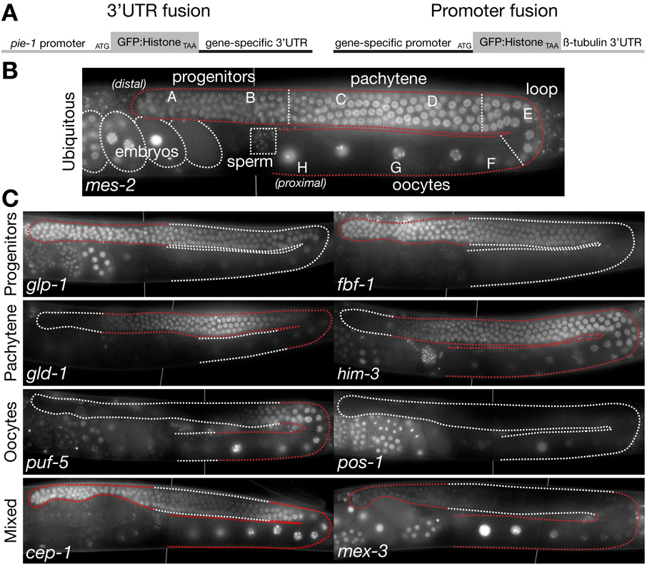
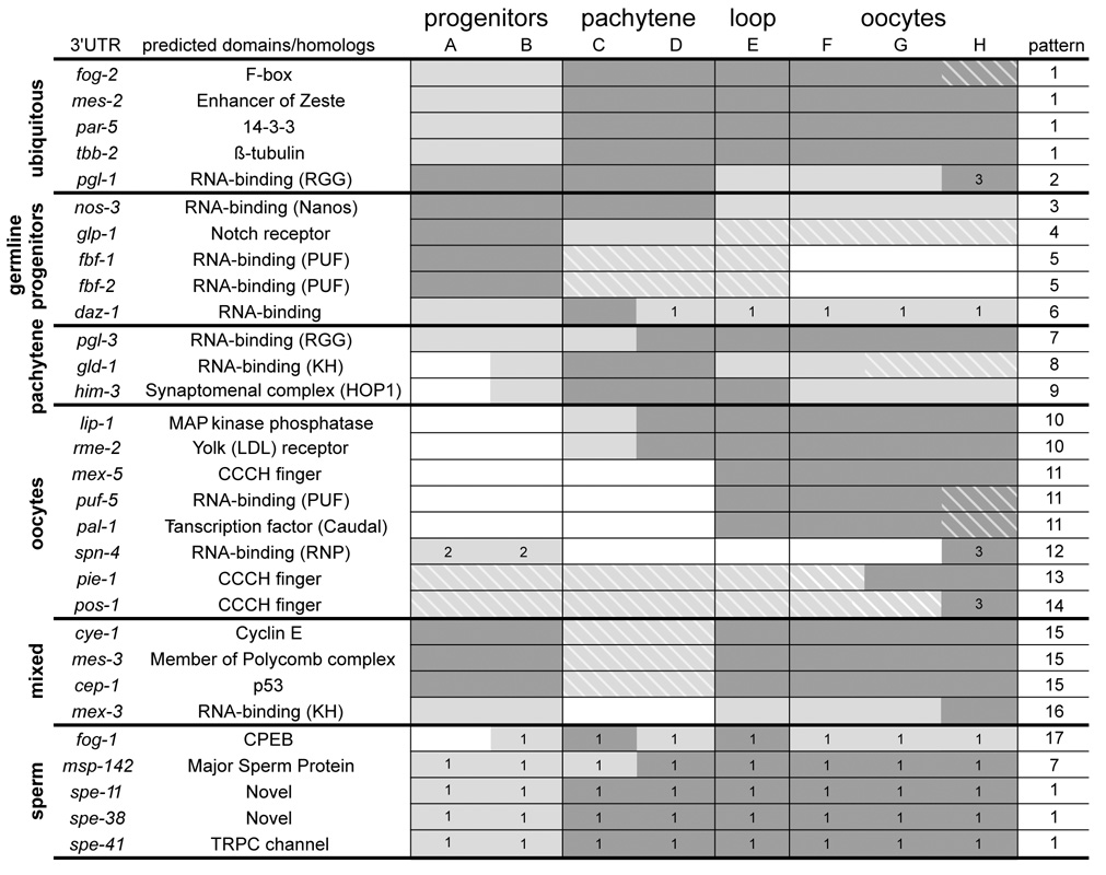
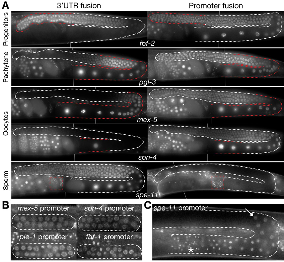
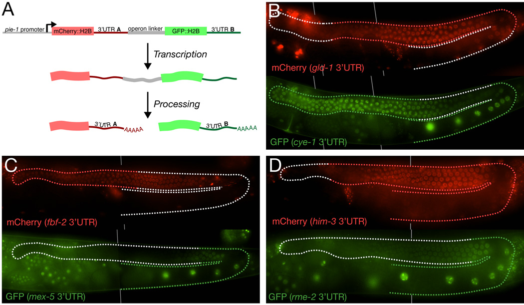
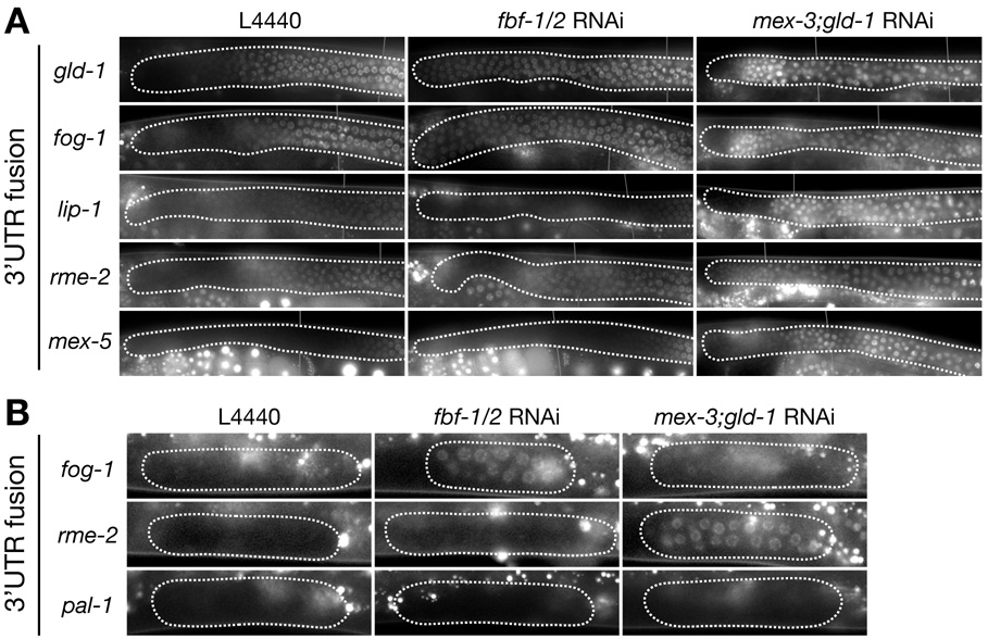
How genes are regulated to produce the correct assortment of proteins for every cell type is a fundamental question in biology. For many genes, regulation begins at the DNA level with the use of promoter sequences to control transcription. Regulation can also occur after transcription using sequences in the 3' untranslated region (UTR) of the mRNA to affect mRNA stability and/or translation [1]. The C. elegans gonad is an excellent tissue to study gene regulation during development: In the adult, germ cells are arranged in order of differentiation, with undifferentiated progenitors at one end of the gonad, cells in meiotic prophase in the middle, and gametes at the other end [2]. Using a transgenic assay, we have compared the contribution of promoters and 3' UTRs to gene regulation during germline development. We find that for most genes tested, 3' UTRs are sufficient for regulation. With the exception of promoters activated during spermatogenesis, promoters are permissive for expression in all germ cell types (from progenitors to oocytes and sperm). In progenitors, 3' UTRs inhibit the production of meiotic and oocyte proteins by posttranscriptional mechanisms involving PUF- and KH-domain RNA-binding proteins. Our findings indicate that many genes rely primarily on 3' UTRs, not promoters, for regulation during germline development.


Figures

Figure 1
Figure 1. 3’UTR fusions in adult hermaphrodites
A. Schematics showing the design of 3’UTR and promoter fusions. Both contain green fluorescent protein (GFP) fused to histone H2B fusion as the protein reporter. H2B associates dynamically with chromatin [31]. Chromatin localization facilitates detection and turnover is fast enough to visualize changes in gene expression even in non-dividing cells ([10]; this study). The pie-1 promoter and β-tubulin 3’ UTR (tbb-2) are permissive for expression in all germ cells. B. Photomicrograph of a single adult gonadal arm (outlined with stippled line) expressing the mes-2 3’UTR fusion (ubiquitous expression). By this stage, hermaphrodites are producing oocytes. Sperm (produced during the L4 larval stage) are stored in the spermatheca. Gonad is divided into different zones used for systematic scoring of expression patterns as in Fig. 2: A is first half of mitotic zone, containing germline stem cells, B is second half of mitotic zone containing cells that have initiated meiotic S phase, C and D are pachytene regions, E is loop region where cells transition from pachytene to diakinesis and initiate oocyte formation, F–H is region of oocyte growth, H denotes last oocyte [1]. C. Photomicrographs as in B showing examples of different 3’ UTR fusions, arranged based on the primary site of protein expression as reported in the literature. For each gene, red stippling outlines the region of the gonad where high levels of endogenous protein have been detected (Sup. Table 1, for references). See Fig. 3 and Sup. Fig.1 for photomicrographs of all other 3’ UTR fusions examined in this study and Fig. 2 for a schematic summary. In this and all subsequent figures, vertical lines are used to indicate where two photomicrographs of the same animal have been merged.
Figure 2
Figure 2. Summary of all 3’UTR fusions examined in this study
Genes are arranged based on the primary site of protein expression as reported in the literature. GFP:H2B expression was scored for each gonadal region (A–H) as defined in Fig. 1B. Dark grey indicates strongest domain(s) of GFP:H2B expression, light grey: weaker domain(s) of GFP expression, and white: no GFP expression. (Evaluation of strong and weak expression was made by comparing GFP intensities directly within individual gonads; at least 20 hermaphrodites were examined for each fusion). Hatching denotes regions where GFP:H2B levels are low/declining and where the endogenous protein has not been reported to be expressed. This type of discrepancy is likely due to perdurance of the H2B fusion or to undetected low levels of endogenous protein. For fog-2, daz-1, gld-1, pal-1, pie-1, and cep-1, replacing H2B with the native ORF gave a pattern more closely matching the endogenous pattern (Sup. Fig. 2). 1. GFP:H2B levels are steady in this region, but endogenous protein has not been reported here. This type of discrepancy suggests that the 3’UTR is insufficient for proper regulation. 5 of 6 3’UTRs in this category come from genes coding for proteins expressed specifically during spermatogenesis. Note that the fog-1 3’UTR is sufficient to confer proper regulation (repression) in germline progenitors, but is insufficient to inhibit expression in germlines undergoing oogenesis. The only other gene in this category is daz-1. DAZ-1 protein has been reported to be expressed in the distal germline (Sup. Table 1), but the daz-1 3’UTR fusion is also expressed in oocytes. The DAZ-1ORF: 3’UTR fusion shows reduced expression in oocytes (Sup. Fig. 2). 2. Whether SPN-4 is expressed or not in this region has not been reported. 3. GFP:H2B expression was most prominent in oocytes undergoing maturation.
Fig. 3
Fig. 3. Comparison of 3’ UTR and promoter fusions
A. Photomicrographs of adult gonads expressing 3’UTR or promoter fusions for the genes indicated. Additional promoter fusions are shown in Sup. Fig. 3. For fbf-2, pgl-3, mex-5 and spn-4, the 3’UTR fusions show the expected cell-type specificity (highest GFP fluorescence matches red stippling), but the promoter fusions do not. In contrast for spe-11, the promoter fusion shows the expected cell type specificity (sperm, see box), but the 3’UTR fusion does not. B and C. Photomicrographs of larval gonadal arms expressing the indicated promoter fusions. All fusions contain the tubulin 3’UTR. B.The mex-5, spn-4, pie-1 and fbf-1 promoter fusions are active in all germline progenitors in L2 gonads. C. In contrast, the spe-11 promoter fusion does not become active until the L4 stage when spermatogenesis begins. GFP:H2B is first detected in late pachytene cells (arrow) and is maintained through mature sperm (asterisk).
Figure 4
Figure 4. 3’UTRs function post-transcriptionally
A. Schematic showing the design of operon transgenes. The operon linker is the intercistronic region from the gpd-2/gpd-3 operon. This 138bp region promotes transplicing and has no promoter activity [32]. B–D. Photomicrographs of adult hermaphrodite gonads expressing the indicated operon transgenes. Top and bottom figures show expression of mCherry and GFP, respectively, in the same animal. Red and green stippling indicated regions of endogenous expression.
Figure 5
Figure 5. Regulation of 3’UTR fusions in the distal gonad by FBF-1/2, MEX-3 and GLD-1
A. Fluorescence photomicrographs of distal arms (A, B and C regions in Fig. 1b) from adult hermaphrodites expressing the indicated 3’ UTR fusions, and fed either control bacteria (L4440) or bacteria expressing double-stranded RNA against fbf-1 and fbf-2 or mex-3 and gld-1. See Sup. Table 2 for numbers and additional 3’ UTR fusions examined. Note that lip-1 has low, but non-zero expression throughout the distal region. B. Photomicrographs of L2 gonadal arms expressing the indicated fusions, and fed either control bacteria (L4440) or bacteria expressing double-stranded RNA against fbf-1 and fbf-2 or mex-3 and gld-1. The RNAi conditions used in these experiments allow the small pool of L2 germline progenitors (~30 cells per arm) to proliferate as in wild-type (~1000 cells per arm by adult stage), indicating that the cells retain mitotic potential and have not yet been transformed. See Sup. Table 3 for numbers and additional 3’UTR fusions examined. Bright foci outside of the gonad are autofluorescent gut granules.

Comment in

Similar articles

Cited by

References

    1. Hubbard EJ, Greenstein D. Introduction to the germ line. WormBook. 2005:1–4. - PMC - PubMed
    1. Ellis R, Schedl T. Sex determination in the germ line. WormBook. 2007:1–13. - PMC - PubMed
    1. Kimble J, Crittenden SL. Germline proliferation and its control. WormBook. 2005:1–14. - PMC - PubMed
    1. Reinke V. Germline genomics. WormBook. 2006:1–10. - PMC - PubMed
    1. Shim YH. elt-1, a gene encoding a Caenorhabditis elegans GATA transcription factor, is highly expressed in the germ lines with msp genes as the potential targets. Mol. Cells. 1999;9:535–541. - PubMed

Publication types