. 2008 Nov 28;283(48):33674-84.
doi: 10.1074/jbc.M804288200. Epub 2008 Sep 24.

Evolutionary differences in glycosaminoglycan fine structure detected by quantitative glycan reductive isotope labeling

Affiliations

Evolutionary differences in glycosaminoglycan fine structure detected by quantitative glycan reductive isotope labeling

Roger Lawrence et al. J Biol Chem. .

Abstract

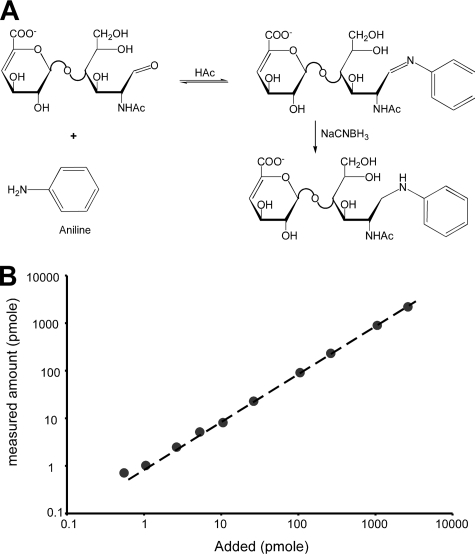
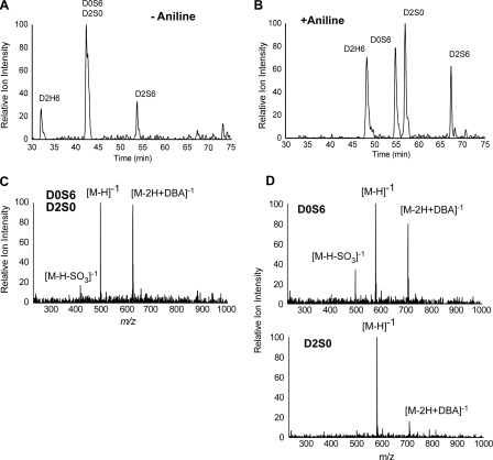
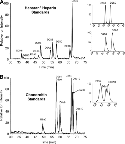
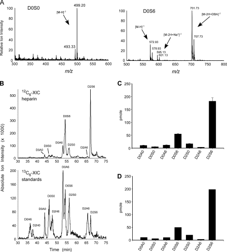
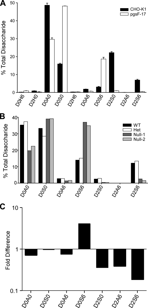
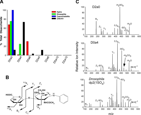
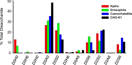
To facilitate qualitative and quantitative analysis of glycosaminoglycans, we tagged the reducing end of lyase-generated disaccharides with aniline-containing stable isotopes (12C6 and 13C6). Because different isotope tags have no effect on chromatographic retention times but can be discriminated by a mass detector, differentially isotope-tagged samples can be compared simultaneously by liquid chromatography/mass spectrometry and quantified by admixture with known amounts of standards. The technique is adaptable to all types of glycosaminoglycans, and its sensitivity is only limited by the type of mass spectrometer available. We validated the method using commercial heparin and keratan sulfate as well as heparan sulfate isolated from mutant and wild-type Chinese hamster ovary cells, and select tissues from mutant and wild-type mice. This new method provides more robust, reliable, and sensitive means of quantitative evaluation of glycosaminoglycan disaccharide compositions than existing techniques allowing us to compare the chondroitin and heparan sulfate compositions of Hydra vulgaris, Drosophila melanogaster, Caenorhabditis elegans, and mammalian cells. Our results demonstrate significant differences in glycosaminoglycan structure among these organisms that might represent evolutionarily distinct functional motifs.


Figures

FIGURE 1.
FIGURE 1.
Aniline-tagging of GAG disaccharides. A, the reducing form of each GAG disaccharide can form a Schiff base with the amine group of aniline. The resulting imine is then reduced with sodium cyanoborohydride resulting in a stable derivative. B, to determine the sensitivity and linear range of GRIL disaccharide analysis, D2S0 was labeled with either [12C6]aniline or [13C6]aniline. Differing amounts of [12C6]aniline-labeled D2S0 (between 0.5 pmol and 1 nmol) were mixed in triplicate with 25 pmol of [13C6]aniline-labeled D2S0 prior to LC/MS analysis. The measured amount (y-axis) is plotted against the amount of [12C6]aniline-labeled disaccharide added (x-axis). The standard error was less than the diameter of the symbols and <5%.
FIGURE 2.
FIGURE 2.
Enhanced detection and resolution of disaccharides. A, the accumulated XIC for disulfated disaccharide ions present in equimolar mixtures of untagged D2H6, D0S6, D2S0, and D2S6 are shown. The accumulative XIC is the summation of individual ionic currents for the free molecular ion, [M-H]-1 (m/z = 496), the adduction ion formed with the ion-pairing reagent, [M-2H+DBA]-1 (m/z = 625), and in-source desulfation of D2S6 [M-H-SO3]-1 (m/z = 496). The two isobaric isomers D0S6 and D2S0 did not resolve. B, the accumulated XIC for D2H6, D0S6, D2S0, and D2S6 tagged with [13C6]aniline is shown. The accumulative XIC is the summation of individual ionic currents for the free molecular ion, [M-H]-1 (m/z = 573), the adduction ion formed with the ion-pairing reagent, [M-2H+DBA]-1 (m/z = 702), and the in-source desulfation of D2S6 [M-H-SO3]-1 (m/z = 573). D0S6 and D2S0, which were not resolved in their underivatized state, are resolved as aniline derivatives. C, corresponding mass spectrum for underivatized D0S6 and D2S0 showing the free molecular ions, [M-H]-1, adduction ions formed with the ion-pairing reagent, [M-2H+DBA]-1, and desulfated in-source fragment ions [M-H-SO3]-1. D, the corresponding mass spectra for D0S6 and D2S0 tagged with [13C6]aniline.
FIGURE 3.
FIGURE 3.
GRIL-LC/MS analysis of aniline-tagged HS and CS disaccharides. A, XIC chromatographs for aniline-tagged HS disaccharide standards resolved by LC/MS. The panels on the right show that the two 3-O-sulfated HS disaccharides D2S3 and D2S9 can also be resolved after aniline tagging as can the two N-acetylated isobaric isomers D0A6 and D2A0. The only standard not resolved was D0H0, which co-elutes with the salt peak, thus suppressing its ionic signature. B, XIC chromatographs for CS disaccharide standards after aniline tagging. The panel on the right expands the region where disulfated CS disaccharide standards elute showing partial separation of D2a6 and D0a10. The two isobaric isomers D0a4 and D2a0 did not separate under these conditions but can be easily distinguished by CID (see Fig. 7). All XIC traces were generated with the appropriate m/z values for the HS and CS disaccharides listed in Table 1.
FIGURE 4.
FIGURE 4.
Qualitative structural analysis of KS GAG chains using GRIL-LC/MS. A, generalized structure of corneal KS I showing the cleavage sites for keratanase (solid arrows) and keratanase II (broken arrows). The symbols used for monosaccharides subunits: sialic acid (purple diamond), galactose (yellow circle), N-acetylglucosamine (blue squares), mannose (green circles), and fucose (red triangle) are as previously described (grtc.ucsd.edu/symbol.html). The presence of 6-O-sulfate is also indicated (6S) above each sulfated monosaccharide. B, bovine corneal KS was digested with either keratanase (“Experimental Procedures”), aniline-tagged and subjected to LC/MS for analysis of digestion products. The chromatographs show the relative ion intensity (XIC) of digestion products eluting from the C18 column. The indicated peak assignments were based on retention time, mass, and the known activity of the keratanases used. The inset in the lower panel shows the elution of two species released by keratanase II digestion with degree of polymerization of 4 and 5 (dp4 and dp5) based on mass. The XIC traces were generated with the appropriate m/z values for KS disaccharides listed in Table 1 and for m/z values for the major expected ions of oligosaccharides released after keratanase digestion. For keratanase, these included: dp4 ([M-3H+DBA]-2, m/z = 596), dp6 ([M-5H+3DBA]-2, m/z = 987), and dp8 ([M-5H+3DBA]-2, m/z = 1250). For keratanase II these included dp4 species g0A6g0A6 ([M-2H]-2, m/z = 491.5), g0A6g6A6 and g6A6g0A6 ([M-3H+DBA]-2, m/z = 596), and g6A6g6A6 ([M-4H+2DBA]-2, m/z = 700.5). The inset XIC trace in the Keratanase II panel was generated for the putative dp5 species Neu5Ac-g0A6g0A6 ([M-2H]-2, m/z = 637), which also reveals a putative minor adduction ion for g6A6g6A6 ([M-3H+DBA]-2, m/z = 636). The mass spectra of the small peak eluting just after 80 min in the upper chromatogram and the small peak eluting after the putative g6A6g6A6 peak in the lower chromatogram do not correspond to any known keratan sulfate-derived oligosaccharides and are most likely contaminants.
FIGURE 5.
FIGURE 5.
GRIL-LC/MS quantitation of porcine intestinal mucosal heparin. Disaccharides derived from heparin were tagged with [12C6]aniline and mixed with [13C6]aniline disaccharide standards (25 pmol each). A, the mass spectra for a representative low abundance disaccharide, D0S0, and a higher abundance disaccharide, D0S6, are shown. Note the 6 atomic mass units difference between the [12C6]aniline-tagged residue and the corresponding [13C6]aniline-tagged standard for the free molecular ions ([M-H]-1), the sodium adducts ([M-2H+Na+]-1), and the ion pairing reagent adducts ([M-2H+DBA]-1). Adduction ions are typically detected for disaccharides with two or more sulfates (compare the mass spectra for D0S0 and D0S6, see Table 1). B, the individual XIC profiles for the [12C6]aniline-tagged sample and [13C6]aniline-tagged standards for one of three separate analyses. The XIC traces were generated using the m/z values for HS disaccharide standards listed in Table 1. C, GRIL-LC/MS determined disaccharide composition of porcine heparin (n = 3), error bars represent the standard error. D, analysis of an identical sample using the anion-exchange high performance liquid chromatography with post column derivatization and fluorescence detection. The preparation of heparin used in this study did not contain measurable amounts of D0H0, D0H6, D2H0, or D2H6 detected by either LC/MS-GRIL or post column derivatization with 2-cyanoacetamide. The rare disaccharide D2A0, which is also detectable by GRIL-LC/MS (Fig. 3A), was not analyzed in this experiment.
FIGURE 6.
FIGURE 6.
GRIL-LC/MS analysis of disaccharides in wild-type and Hs2st-deficient mutant cells and mice. A, the disaccharide profile for HS isolated from wild-type CHO-K1 cells and from pgsF-17 mutant cells (Hs2st-deficient), error bars represent the standard error of the means (n = 3). The results are reported as percentage of total disaccharide. B, the disaccharide profile for HS isolated from Hs2st-/-, Hs2st+/-, or wild-type mouse embryos. The results are reported as percentage of total disaccharide recovered from each sample. C, the disaccharides derived from HS isolated from the liver of Hs2stf/fAlbCre+ and Hs2stf/fAlbCre- mice were tagged with [13C6]aniline or [12C6]aniline, respectively. The samples were mixed and then analyzed by LC/MS. The ratio of 13C and 12C recovered for each disaccharide is shown.
FIGURE 7.
FIGURE 7.
GRIL-LC/MS disaccharide analysis of CS isolated from Hydra, nematodes, and arthropods. A, chondroitins from H. vulgaris, D. melanogaster, C. elegans, and CHO-K1 cells was enzymatically depolymerized and tagged with [12C6]aniline. An equimolar mixture (25 pmol) of [13C6]aniline-tagged CS/DS disaccharide standards was added prior to LC/MS analysis. Results are shown as the percentage of total disaccharides recovered. B, schematic showing possible tandem mass spectra intra-and inter-ring cleavages of aniline-conjugated D0a4, using the nomenclature proposed by Domon and Costello (49). C, tandem mass spectra of D2a0, D0a4, and the uncharacterized monosulfated disaccharide (Drosophila dp2(1SO4)) present in the D. melanogaster chondroitin sample.
FIGURE 8.
FIGURE 8.
GRIL-LC/MS disaccharide analysis of HS from Hydra, nematodes, and arthropods. HS samples were depolymerized by exhaustive digestion with a mixture of heparan lyases, reacted with propionic anhydride, tagged with [12C6]aniline, and mixed with [13C6]aniline-tagged HS disaccharide standards. The proportion of each disaccharide in the samples was then determined by comparison to the standards, including [13C6]aniline-tagged standards for propionylated N-unsubstituted disaccharides. Significant levels of D0H0 were detected in H. vulgaris and D. melanogaster HS but not in C. elegans or CHO cells.

References

    1. Kinoshita-Toyoda, A., Yamada, S., Haslam, S. M., Khoo, K. H., Sugiura, M., Morris, H. R., Dell, A., and Sugahara, K. (2004) Biochemistry 43 11063-11074 - PubMed
    1. Esko, J. D., and Selleck, S. B. (2002) Annu. Rev. Biochem. 71 435-471 - PubMed
    1. Toyoda, H., Kinoshita-Toyoda, A., and Selleck, S. B. (2000) J. Biol. Chem. 275 2269-2275 - PubMed
    1. Toyoda, H., Kinoshita-Toyoda, A., Fox, B., and Selleck, S. B. (2000) J. Biol. Chem. 275 21856-21861 - PubMed
    1. Yamada, S., Morimoto, H., Fujisawa, T., and Sugahara, K. (2007) Glycobiology 17 886-894 - PubMed

Publication types

LinkOut - more resources