. 2008 Dec 12;283(50):34696-703.
doi: 10.1074/jbc.M802210200. Epub 2008 Sep 25.

Ziram causes dopaminergic cell damage by inhibiting E1 ligase of the proteasome

Affiliations

Ziram causes dopaminergic cell damage by inhibiting E1 ligase of the proteasome

Arthur P Chou et al. J Biol Chem. .

Abstract

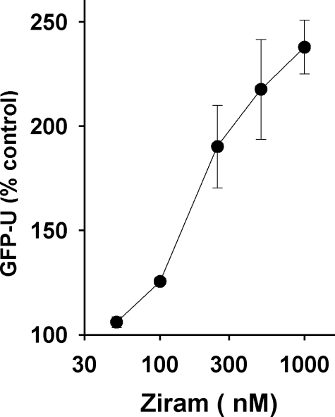
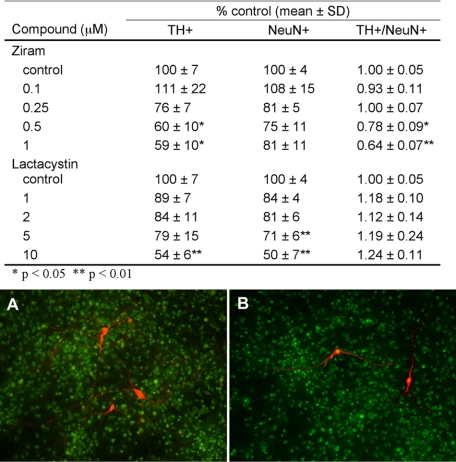
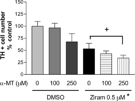
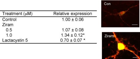
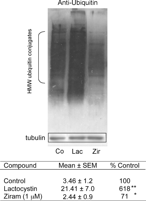
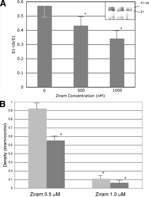
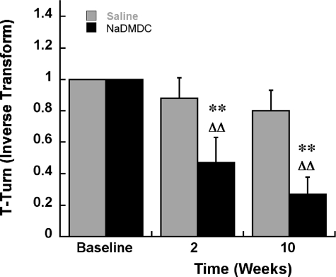
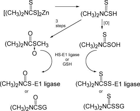
The etiology of Parkinson disease (PD) is unclear but may involve environmental toxins such as pesticides leading to dysfunction of the ubiquitin proteasome system (UPS). Here, we measured the relative toxicity of ziram (a UPS inhibitor) and analogs to dopaminergic neurons and examined the mechanism of cell death. UPS (26 S) activity was measured in cell lines after exposure to ziram and related compounds. Dimethyl- and diethyldithiocarbamates including ziram were potent UPS inhibitors. Primary ventral mesencephalic cultures were exposed to ziram, and cell toxicity was assessed by staining for tyrosine hydroxylase (TH) and NeuN antigen. Ziram caused a preferential damage to TH+ neurons and elevated alpha-synuclein levels but did not increase aggregate formation. Mechanistically, ziram altered UPS function through interfering with the targeting of substrates by inhibiting ubiquitin E1 ligase. Sodium dimethyldithiocarbamate administered to mice for 2 weeks resulted in persistent motor deficits and a mild reduction in striatal TH staining but no nigral cell loss. These results demonstrate that ziram causes selective dopaminergic cell damage in vitro by inhibiting an important degradative pathway implicated in the etiology of PD. Chronic exposure to widely used dithiocarbamate fungicides may contribute to the development of PD, and elucidation of its mechanism would identify a new potential therapeutic target.


Figures

FIGURE 1.
FIGURE 1.
Ziram inhibits UPS in HEK cells with 24-h incubation prior to analysis by flow cytometry. The IC50 for ziram was 161 nm.
FIGURE 2.
FIGURE 2.
Ziram- and lactacystin-induced tyrosine hydroxylase cell damage in primary mesencephalic cultures. TH+ neurons were selectively vulnerable to ziram-but not lactacystin-induced damage. Shown below are representative photomicrographs of TH-(red) and NeuN-stained (green) mesencephalic cultures. A, control. B, ziram (1 μm).
FIGURE 3.
FIGURE 3.
Inhibition of dopamine synthesis by α-methyl-l-tyrosine did not attenuate ziram-induced dopamine cell death (n = 14–44 wells per condition). *, p ≤ 0.05, ziram versus dimethyl sulfoxide (DMSO) control. +, not significant.
FIGURE 4.
FIGURE 4.
Ziram increases and lactacystin decreases α-synuclein levels in TH+ neurons. The cells were exposed for 48 h prior to analysis. The relative intensities were measured in a blinded manner in cells that were selected randomly (n = 21–97 cells/condition). Representative α-synuclein-stained cells are shown on the right. Scale bars, 10 microns. **, p ≤ 0.01. Con, control.
FIGURE 5.
FIGURE 5.
Anti-ubiquitin Western blot analysis of lysates from cells following treatment with 5 μm lactacystin, 0. 1 μm rotenone, or 1 μm ziram. The relative densities were determined using the NIH Image program, normalized to tubulin immuoreactivity in the same blot, and expressed as percentages of untreated controls (n = 4). 20 S inhibition using lactacystin resulted in an increase in high molecular weight ubiquitin conjugates, whereas ziram treatment reduced them. **, p ≤ 0.01; *, p ≤ 0.05.
FIGURE 6.
FIGURE 6.
Effect of ziram on E1-ubiquitin conjugates and E1 ligase activity. A, endogenous E1-ubiquitin conjugates in lysates of ziram-treated HEK cells. Ziram resulted in a dose-dependent reduction in the E1-Ub/E1 ratio compared with untreated controls. The Western blot is shown in the inset. B, E1 ligase activity was inhibited by ziram in purified preparations after 5 and 10 min of incubation (n = 4, p ≤ 0.05).
FIGURE 7.
FIGURE 7.
Motor deficits in mice determined by the pole test following 2 weeks of subcutaneous minipump treatment with NaDMDC at 50 mg/kg/day followed by an 8-week post-treatment period (10 weeks from the beginning of the experiment). Inverse Transform, reciprocal of the original data point. Baseline, 1 because untreated mice turn in less than 1 s. **, p < 0.01 compared with saline-treated mice at the same time point. ΔΔ represents p < 0.01 compared with a base line within the same treatment group (2 × 3 Mixed design ANOVA, Fisher's LSD).
FIGURE 8.
FIGURE 8.
Alternative mechanisms for ziram oxidative activation and reaction with 26 S E1 ligase or GSH.

References

    1. Ascherio, A., Chen, H., Weisskopf, M. G., O'Reilly, E., McCullough, M. L., Calle, E. E., Schwarzschild, M. A., and Thun, M. J. (2006) Ann. Neurol. 60197 –203 - PubMed
    1. Kamel, F., Tanner, C., Umbach, D., Hoppin, J., Alavanja, M., Blair, A., Comyns, K., Goldman, S., Korell, M., Langston, J., Ross, G., and Sandler, D. (2007) Am. J. Epidemiol. 165364 –374 - PubMed
    1. Brown, T. P., Rumsby, P. C., Capleton, A. C., Rushton, L., and Levy, L. S. (2006) Environ. Health Perspect. 114156 –164 - PMC - PubMed
    1. McNaught, K. S., and Jenner, P. (2001) Neurosci. Lett. 297191 –194 - PubMed
    1. Manning-Bog, A. B., Reaney, S. H., Chou, V. P., Johnston, L. C., McCormack, A. L., Johnston, J., Langston, J. W., and Di Monte, D. A. (2006) Ann. Neurol. 60256 –260 - PubMed

Publication types

LinkOut - more resources